

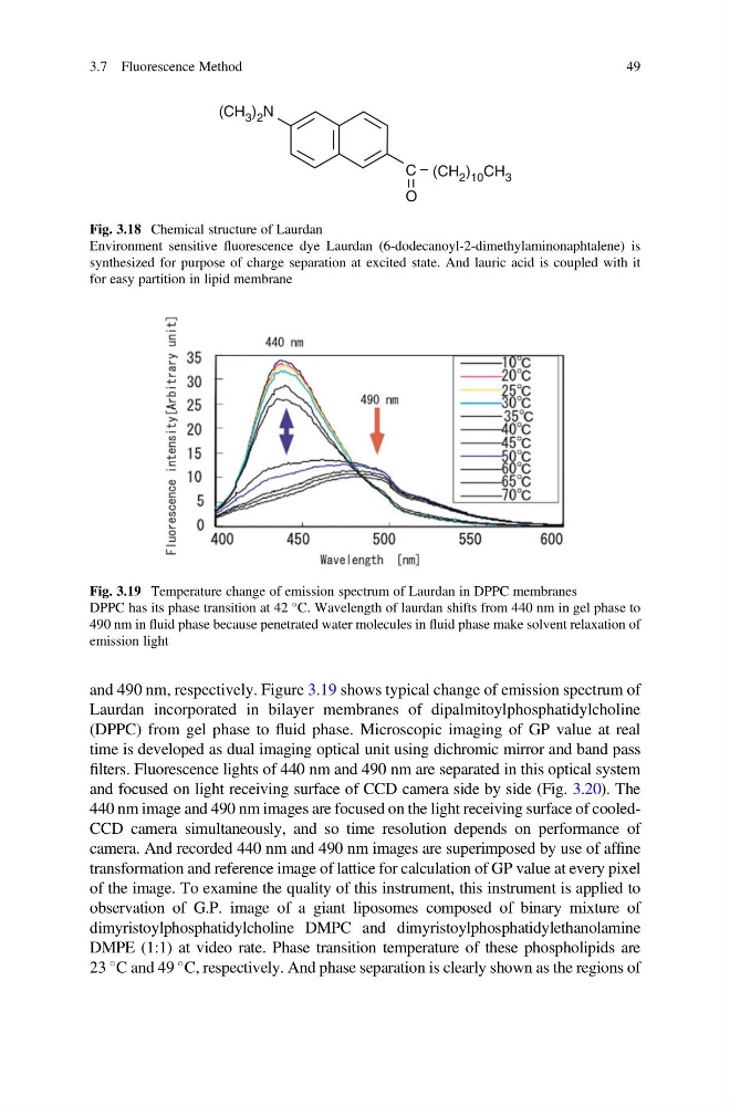
and 490 nm, respectively. Figure 3.19 shows typical change of emission spectrum of
Laurdan incorporated in bilayer membranes of dipalmitoylphosphatidylcholine
(DPPC) from gel phase to fluid phase. Microscopic imaging of GP value at real
time is developed as dual imaging optical unit using dichromic mirror and band pass
filters. Fluorescence lights of 440 nm and 490 nm are separated in this optical system
and focused on light receiving surface of CCD camera side by side (Fig. 3.20). The
440 nm image and 490 nm images are focused on the light receiving surface of cooledCCD camera simultaneously, and so time resolution depends on performance of
camera. And recorded 440 nm and 490 nm images are superimposed by use of affine
transformation and reference image of lattice for calculation of GP value at every pixel
of the image. To examine the quality of this instrument, this instrument is applied to
observation of G.P. image of a giant liposomes composed of binary mixture of
dimyristoylphosphatidylcholine DMPC and dimyristoylphosphatidylethanolamine
DMPE (1:1) at video rate. Phase transition temperature of these phospholipids are
23
C and 49
C, respectively. And phase separation is clearly shown as the regions of
Fig. 3.19 Temperature change of emission spectrum of Laurdan in DPPC membranes
DPPC has its phase transition at 42
C. Wavelength of laurdan shifts from 440 nm in gel phase to
490 nm in fluid phase because penetrated water molecules in fluid phase make solvent relaxation of
emission light
O
(CH 2 ) 10 CH 3
(CH 3 ) 2 N
C
Fig. 3.18 Chemical structure of Laurdan
Environment sensitive fluorescence dye Laurdan (6-dodecanoyl-2-dimethylaminonaphtalene) is
synthesized for purpose of charge separation at excited state. And lauric acid is coupled with it
for easy partition in lipid membrane
3.7 Fluorescence Method
49
Laurdan incorporated in bilayer membranes of dipalmitoylphosphatidylcholine
(DPPC) from gel phase to fluid phase. Microscopic imaging of GP value at real
time is developed as dual imaging optical unit using dichromic mirror and band pass
filters. Fluorescence lights of 440 nm and 490 nm are separated in this optical system
and focused on light receiving surface of CCD camera side by side (Fig. 3.20). The
440 nm image and 490 nm images are focused on the light receiving surface of cooledCCD camera simultaneously, and so time resolution depends on performance of
camera. And recorded 440 nm and 490 nm images are superimposed by use of affine
transformation and reference image of lattice for calculation of GP value at every pixel
of the image. To examine the quality of this instrument, this instrument is applied to
observation of G.P. image of a giant liposomes composed of binary mixture of
dimyristoylphosphatidylcholine DMPC and dimyristoylphosphatidylethanolamine
DMPE (1:1) at video rate. Phase transition temperature of these phospholipids are
23
C and 49
C, respectively. And phase separation is clearly shown as the regions of
Fig. 3.19 Temperature change of emission spectrum of Laurdan in DPPC membranes
DPPC has its phase transition at 42
C. Wavelength of laurdan shifts from 440 nm in gel phase to
490 nm in fluid phase because penetrated water molecules in fluid phase make solvent relaxation of
emission light
O
(CH 2 ) 10 CH 3
(CH 3 ) 2 N
C
Fig. 3.18 Chemical structure of Laurdan
Environment sensitive fluorescence dye Laurdan (6-dodecanoyl-2-dimethylaminonaphtalene) is
synthesized for purpose of charge separation at excited state. And lauric acid is coupled with it
for easy partition in lipid membrane
3.7 Fluorescence Method
49