

3.7.3 Fluorescence Resonance Energy Transfer (FRET)
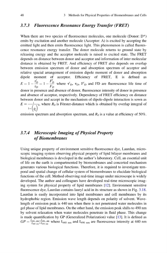
When there are two species of fluorescence molecules, one molecule (Donor: D*)
emits by excitation and another molecule (Acceptor: A) is excited by accepting the
emitted light and then emits fluorescence light. This phenomenon is called fluorescence resonance energy transfer. The donor molecule returns to ground state by
releasing energy and the acceptor molecule is raised to excited state. The FRET
depends on distance between donor and acceptor and information of inter molecular
distance is obtained by FRET. And efficiency of FRET also depends on overlap
between emission spectrum of donor and absorption spectrum of acceptor and
relative spacial arrangement of emission dipole moment of donor and absorption
dipole moment of acceptor. Efficiency of FRET, E is defined as
E ¼ 1 À
τ
0
D
τ D
¼ 1 À
F
0
D
F D
where τ
0
D , τ D , F
0
D and FD are fluorescence life time of
donor in presence and absence of donor, fluorescence intensity of donor in presence
and absence of acceptor, respectively. Dependency of FRET efficiency on distance
between donor and accept in the mechanism of dipole-dipole interaction is sown as
E ¼
1
1þ
r
R 0
6 , where R 0 is Förster-distance which is obtained by overlap integral of
emission spectrum and absorption spectrum, and R 0 is a value at efficiency of 50%.
3.7.4 Microscopic Imaging of Physical Property
of Biomembranes
Using unique property of environment sensitive fluorescence dye, Laurdan, microscopic imaging system observing physical property of lipid bilayer membranes and
biological membranes is developed in the author’s laboratory. Cell, an essential unit
of life on the earth is compartmented by biomembranes and concerted mechanism
generates various biological functions. Therefore, it is required to investigate temporal and spatial change of cellular system of biomembranes to elucidate biological
functions of the cell. Method observing real-time image under microscope is widely
developed. The author and colleagues have developed real-time microscopic imaging system for physical property of lipid membranes [12]. Environment sensitive
fluorescence dye, Laurdan contains lauryl acid in its structure as shown in Fig. 3.18.
Laurdan is easily incorporated into lipid membranes and cell membranes by its
hydrophobic region. Emission wave length depends on polarity of solvent. Wavelength of emission peak is 440 nm when there is not penetrated water molecules in
gel phase of lipid membranes. On the other hand, the emission peak shifts to 490 nm
by solvent relaxation when water molecules penetrate in fluid phase. This change
is made quantification by GP (Generalized Polarization) value [13]. It is defined as
GP ¼
I 440 nm ÀI 490 nm
I 440 nm þI 490 nm
where I 440 nm and I 490 nm are fluorescence intensity at 440 nm
48
3 Methods for Physical Properties of Biomembranes and Cells
When there are two species of fluorescence molecules, one molecule (Donor: D*)
emits by excitation and another molecule (Acceptor: A) is excited by accepting the
emitted light and then emits fluorescence light. This phenomenon is called fluorescence resonance energy transfer. The donor molecule returns to ground state by
releasing energy and the acceptor molecule is raised to excited state. The FRET
depends on distance between donor and acceptor and information of inter molecular
distance is obtained by FRET. And efficiency of FRET also depends on overlap
between emission spectrum of donor and absorption spectrum of acceptor and
relative spacial arrangement of emission dipole moment of donor and absorption
dipole moment of acceptor. Efficiency of FRET, E is defined as
E ¼ 1 À
τ
0
D
τ D
¼ 1 À
F
0
D
F D
where τ
0
D , τ D , F
0
D and FD are fluorescence life time of
donor in presence and absence of donor, fluorescence intensity of donor in presence
and absence of acceptor, respectively. Dependency of FRET efficiency on distance
between donor and accept in the mechanism of dipole-dipole interaction is sown as
E ¼
1
1þ
r
R 0
6 , where R 0 is Förster-distance which is obtained by overlap integral of
emission spectrum and absorption spectrum, and R 0 is a value at efficiency of 50%.
3.7.4 Microscopic Imaging of Physical Property
of Biomembranes
Using unique property of environment sensitive fluorescence dye, Laurdan, microscopic imaging system observing physical property of lipid bilayer membranes and
biological membranes is developed in the author’s laboratory. Cell, an essential unit
of life on the earth is compartmented by biomembranes and concerted mechanism
generates various biological functions. Therefore, it is required to investigate temporal and spatial change of cellular system of biomembranes to elucidate biological
functions of the cell. Method observing real-time image under microscope is widely
developed. The author and colleagues have developed real-time microscopic imaging system for physical property of lipid membranes [12]. Environment sensitive
fluorescence dye, Laurdan contains lauryl acid in its structure as shown in Fig. 3.18.
Laurdan is easily incorporated into lipid membranes and cell membranes by its
hydrophobic region. Emission wave length depends on polarity of solvent. Wavelength of emission peak is 440 nm when there is not penetrated water molecules in
gel phase of lipid membranes. On the other hand, the emission peak shifts to 490 nm
by solvent relaxation when water molecules penetrate in fluid phase. This change
is made quantification by GP (Generalized Polarization) value [13]. It is defined as
GP ¼
I 440 nm ÀI 490 nm
I 440 nm þI 490 nm
where I 440 nm and I 490 nm are fluorescence intensity at 440 nm
48
3 Methods for Physical Properties of Biomembranes and Cells