

3.7.2 Fluorescence Photo-Bleaching Recovery (FPR)
Photo-bleaching recovery is developed for method to obtain diffusion constant
directly [11]. Molecule labeled with fluorescence dye on cell membrane is observed
by fluorescence microscope with weak laser light, and then photo-bleached by strong
laser beam. After then the molecules with fluorescence dyes diffusing into the
bleached spot from area surround the spot is observed with weak laser light for
excitation. Diffusion constant of the molecule labeled with fluorescence dye is
obtained by this temporal observation of inlet molecules. Assuming quenching
process of fluorescence dye is proportional to rate constant αI(r) of simple
non-reversible linear reaction, concentration change of fluorescence dye of
unbleached ones under condition of no transportation is shown as
dc r;t
ð Þ
dt ¼ ÀαI r
ð Þc
r; t
ð Þ where αI(r), r and t are intensity of bleaching light at position r, position and
time of observation, respectively. Differential equation of one species of molecule
transported laterally is shown as
∂c r;t
ð Þ
∂t
¼ D∇
2 c r; t
ð Þ À V 0
∂c r;t
ð Þ
∂x
h
i
where D and V 0
are diffusion constant and velocity of homogenous flow of x-direction, respectively.
Solution of this differential equation under boundary condition c(1, t) ¼ C 0 (homogeneous distribution of fluorescence dye) and initial condition c
(r, 0) ¼ C 0 exp [ÀαTI(r)]: is made curve fitting to process of fluorescence recovery
to obtain diffusion constant.
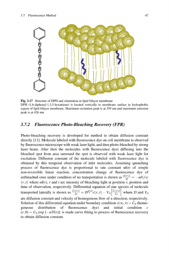
Fig. 3.17 Structure of DPH and orientation in lipid bilayer membrane
DPH (1,6-diphenyl-1,3,5-hexatriene) is located vertically to membrane surface in hydrophobic
region of lipid bilayer membrane. Maximum excitation peak is at 359 nm and maximum emission
peak is at 426 nm
3.7 Fluorescence Method
47
Photo-bleaching recovery is developed for method to obtain diffusion constant
directly [11]. Molecule labeled with fluorescence dye on cell membrane is observed
by fluorescence microscope with weak laser light, and then photo-bleached by strong
laser beam. After then the molecules with fluorescence dyes diffusing into the
bleached spot from area surround the spot is observed with weak laser light for
excitation. Diffusion constant of the molecule labeled with fluorescence dye is
obtained by this temporal observation of inlet molecules. Assuming quenching
process of fluorescence dye is proportional to rate constant αI(r) of simple
non-reversible linear reaction, concentration change of fluorescence dye of
unbleached ones under condition of no transportation is shown as
dc r;t
ð Þ
dt ¼ ÀαI r
ð Þc
r; t
ð Þ where αI(r), r and t are intensity of bleaching light at position r, position and
time of observation, respectively. Differential equation of one species of molecule
transported laterally is shown as
∂c r;t
ð Þ
∂t
¼ D∇
2 c r; t
ð Þ À V 0
∂c r;t
ð Þ
∂x
h
i
where D and V 0
are diffusion constant and velocity of homogenous flow of x-direction, respectively.
Solution of this differential equation under boundary condition c(1, t) ¼ C 0 (homogeneous distribution of fluorescence dye) and initial condition c
(r, 0) ¼ C 0 exp [ÀαTI(r)]: is made curve fitting to process of fluorescence recovery
to obtain diffusion constant.
Fig. 3.17 Structure of DPH and orientation in lipid bilayer membrane
DPH (1,6-diphenyl-1,3,5-hexatriene) is located vertically to membrane surface in hydrophobic
region of lipid bilayer membrane. Maximum excitation peak is at 359 nm and maximum emission
peak is at 426 nm
3.7 Fluorescence Method
47