

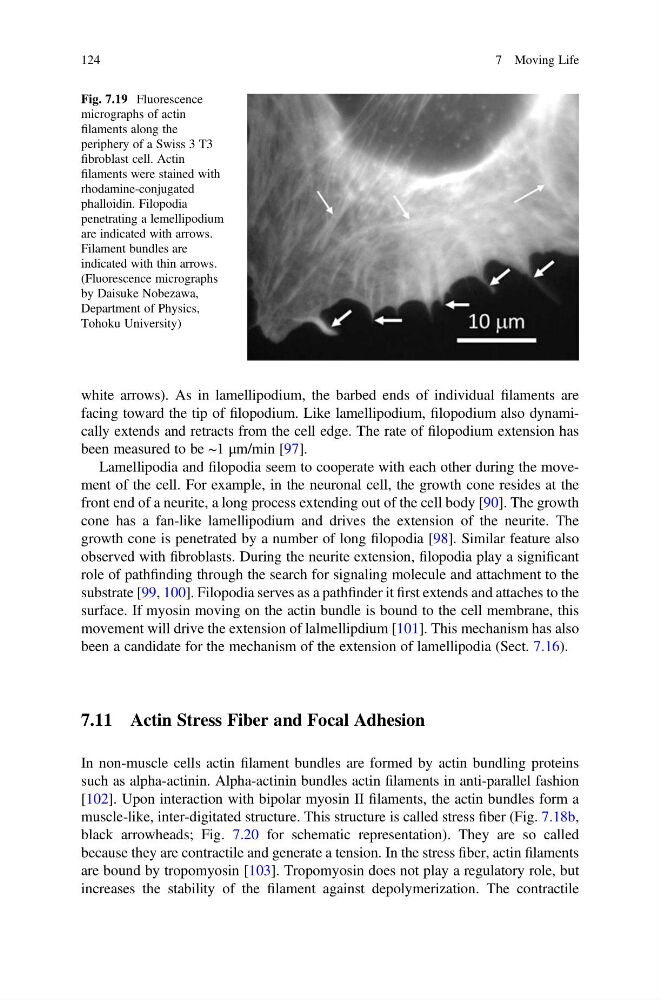
white arrows). As in lamellipodium, the barbed ends of individual filaments are
facing toward the tip of filopodium. Like lamellipodium, filopodium also dynamically extends and retracts from the cell edge. The rate of filopodium extension has
been measured to be ~1 μm/min [97].
Lamellipodia and filopodia seem to cooperate with each other during the movement of the cell. For example, in the neuronal cell, the growth cone resides at the
front end of a neurite, a long process extending out of the cell body [90]. The growth
cone has a fan-like lamellipodium and drives the extension of the neurite. The
growth cone is penetrated by a number of long filopodia [98]. Similar feature also
observed with fibroblasts. During the neurite extension, filopodia play a significant
role of pathfinding through the search for signaling molecule and attachment to the
substrate [99, 100]. Filopodia serves as a pathfinder it first extends and attaches to the
surface. If myosin moving on the actin bundle is bound to the cell membrane, this
movement will drive the extension of lalmellipdium [101]. This mechanism has also
been a candidate for the mechanism of the extension of lamellipodia (Sect. 7.16).
7.11 Actin Stress Fiber and Focal Adhesion
In non-muscle cells actin filament bundles are formed by actin bundling proteins
such as alpha-actinin. Alpha-actinin bundles actin filaments in anti-parallel fashion
[102]. Upon interaction with bipolar myosin II filaments, the actin bundles form a
muscle-like, inter-digitated structure. This structure is called stress fiber (Fig. 7.18b,
black arrowheads; Fig. 7.20 for schematic representation). They are so called
because they are contractile and generate a tension. In the stress fiber, actin filaments
are bound by tropomyosin [103]. Tropomyosin does not play a regulatory role, but
increases the stability of the filament against depolymerization. The contractile
Fig. 7.19 Fluorescence
micrographs of actin
filaments along the
periphery of a Swiss 3 T3
fibroblast cell. Actin
filaments were stained with
rhodamine-conjugated
phalloidin. Filopodia
penetrating a lemellipodium
are indicated with arrows.
Filament bundles are
indicated with thin arrows.
(Fluorescence micrographs
by Daisuke Nobezawa,
Department of Physics,
Tohoku University)
124
7 Moving Life
facing toward the tip of filopodium. Like lamellipodium, filopodium also dynamically extends and retracts from the cell edge. The rate of filopodium extension has
been measured to be ~1 μm/min [97].
Lamellipodia and filopodia seem to cooperate with each other during the movement of the cell. For example, in the neuronal cell, the growth cone resides at the
front end of a neurite, a long process extending out of the cell body [90]. The growth
cone has a fan-like lamellipodium and drives the extension of the neurite. The
growth cone is penetrated by a number of long filopodia [98]. Similar feature also
observed with fibroblasts. During the neurite extension, filopodia play a significant
role of pathfinding through the search for signaling molecule and attachment to the
substrate [99, 100]. Filopodia serves as a pathfinder it first extends and attaches to the
surface. If myosin moving on the actin bundle is bound to the cell membrane, this
movement will drive the extension of lalmellipdium [101]. This mechanism has also
been a candidate for the mechanism of the extension of lamellipodia (Sect. 7.16).
7.11 Actin Stress Fiber and Focal Adhesion
In non-muscle cells actin filament bundles are formed by actin bundling proteins
such as alpha-actinin. Alpha-actinin bundles actin filaments in anti-parallel fashion
[102]. Upon interaction with bipolar myosin II filaments, the actin bundles form a
muscle-like, inter-digitated structure. This structure is called stress fiber (Fig. 7.18b,
black arrowheads; Fig. 7.20 for schematic representation). They are so called
because they are contractile and generate a tension. In the stress fiber, actin filaments
are bound by tropomyosin [103]. Tropomyosin does not play a regulatory role, but
increases the stability of the filament against depolymerization. The contractile
Fig. 7.19 Fluorescence
micrographs of actin
filaments along the
periphery of a Swiss 3 T3
fibroblast cell. Actin
filaments were stained with
rhodamine-conjugated
phalloidin. Filopodia
penetrating a lemellipodium
are indicated with arrows.
Filament bundles are
indicated with thin arrows.
(Fluorescence micrographs
by Daisuke Nobezawa,
Department of Physics,
Tohoku University)
124
7 Moving Life