

7.10 Lamllipodia and Filopodia: Protrusive Membranous
Structures of the Cell
The cell suspended in solution and not attached to a surface is spherical, because of
the lateral tension in the plasma membrane [94], but the adhered cell spreads on the
surface attaining flat morphology. In the initial stage of the spreading, the cell
assumes isotropic, round shape. Later, cell shape becomes more polarized
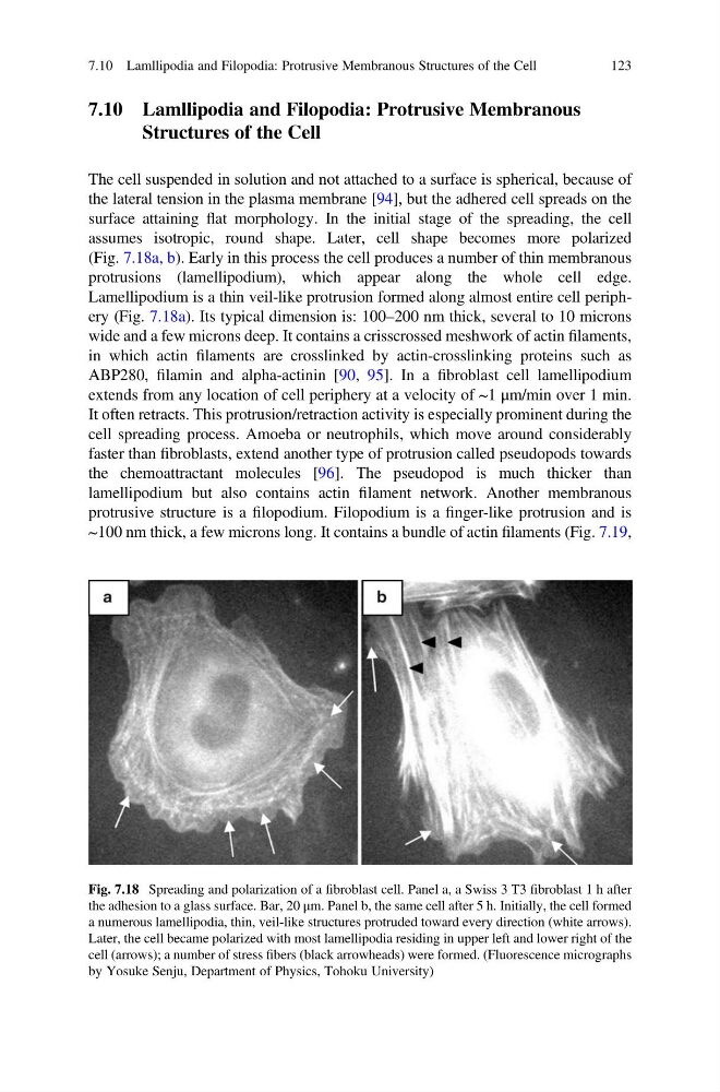
(Fig. 7.18a, b). Early in this process the cell produces a number of thin membranous
protrusions (lamellipodium), which appear along the whole cell edge.
Lamellipodium is a thin veil-like protrusion formed along almost entire cell periphery (Fig. 7.18a). Its typical dimension is: 100–200 nm thick, several to 10 microns
wide and a few microns deep. It contains a crisscrossed meshwork of actin filaments,
in which actin filaments are crosslinked by actin-crosslinking proteins such as
ABP280, filamin and alpha-actinin [90, 95]. In a fibroblast cell lamellipodium
extends from any location of cell periphery at a velocity of ~1 μm/min over 1 min.
It often retracts. This protrusion/retraction activity is especially prominent during the
cell spreading process. Amoeba or neutrophils, which move around considerably
faster than fibroblasts, extend another type of protrusion called pseudopods towards
the chemoattractant molecules [96]. The pseudopod is much thicker than
lamellipodium but also contains actin filament network. Another membranous
protrusive structure is a filopodium. Filopodium is a finger-like protrusion and is
~100 nm thick, a few microns long. It contains a bundle of actin filaments (Fig. 7.19,
Fig. 7.18 Spreading and polarization of a fibroblast cell. Panel a, a Swiss 3 T3 fibroblast 1 h after
the adhesion to a glass surface. Bar, 20 μm. Panel b, the same cell after 5 h. Initially, the cell formed
a numerous lamellipodia, thin, veil-like structures protruded toward every direction (white arrows).
Later, the cell became polarized with most lamellipodia residing in upper left and lower right of the
cell (arrows); a number of stress fibers (black arrowheads) were formed. (Fluorescence micrographs
by Yosuke Senju, Department of Physics, Tohoku University)
7.10 Lamllipodia and Filopodia: Protrusive Membranous Structures of the Cell
123
Structures of the Cell
The cell suspended in solution and not attached to a surface is spherical, because of
the lateral tension in the plasma membrane [94], but the adhered cell spreads on the
surface attaining flat morphology. In the initial stage of the spreading, the cell
assumes isotropic, round shape. Later, cell shape becomes more polarized
(Fig. 7.18a, b). Early in this process the cell produces a number of thin membranous
protrusions (lamellipodium), which appear along the whole cell edge.
Lamellipodium is a thin veil-like protrusion formed along almost entire cell periphery (Fig. 7.18a). Its typical dimension is: 100–200 nm thick, several to 10 microns
wide and a few microns deep. It contains a crisscrossed meshwork of actin filaments,
in which actin filaments are crosslinked by actin-crosslinking proteins such as
ABP280, filamin and alpha-actinin [90, 95]. In a fibroblast cell lamellipodium
extends from any location of cell periphery at a velocity of ~1 μm/min over 1 min.
It often retracts. This protrusion/retraction activity is especially prominent during the
cell spreading process. Amoeba or neutrophils, which move around considerably
faster than fibroblasts, extend another type of protrusion called pseudopods towards
the chemoattractant molecules [96]. The pseudopod is much thicker than
lamellipodium but also contains actin filament network. Another membranous
protrusive structure is a filopodium. Filopodium is a finger-like protrusion and is
~100 nm thick, a few microns long. It contains a bundle of actin filaments (Fig. 7.19,
Fig. 7.18 Spreading and polarization of a fibroblast cell. Panel a, a Swiss 3 T3 fibroblast 1 h after
the adhesion to a glass surface. Bar, 20 μm. Panel b, the same cell after 5 h. Initially, the cell formed
a numerous lamellipodia, thin, veil-like structures protruded toward every direction (white arrows).
Later, the cell became polarized with most lamellipodia residing in upper left and lower right of the
cell (arrows); a number of stress fibers (black arrowheads) were formed. (Fluorescence micrographs
by Yosuke Senju, Department of Physics, Tohoku University)
7.10 Lamllipodia and Filopodia: Protrusive Membranous Structures of the Cell
123