

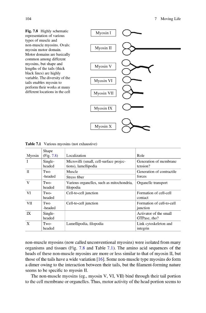
non-muscle myosins (now called unconventional myosins) were isolated from many
organisms and tissues (Fig. 7.8 and Table 7.1). The amino acid sequences of the
heads of these non-muscle myosins are more or less similar to that of myosin II, but
those of the tails have a wide variation [16]. Some non-muscle type myosins do form
a dimer owing to the interaction between their tails, but the filament-forming nature
seems to be specific to myosin II.
The non-muscle myosins (eg., myosin V, VI, VII) bind through their tail portion
to the cell membrane or organelles. Thus, motor activity of the head portion seems to
Myosin I
Myosin II
Myosin V
Myosin VI
Myosin VII
Myosin IX
Myosin X
Fig. 7.8 Highly schematic
representation of various
types of muscle and
non-muscle myosins. Ovals:
myosin motor domain.
Motor domains are basically
common among different
myosins, but shape and
lengths of the tails (thick
black lines) are highly
variable. The diversity of the
tails enables myosin to
perform their works at many
different locations in the cell
Table 7.1 Various myosins (not exhaustive)
Myosin
Shape
(Fig. 7.8)
Localization
Role
I
Singleheaded
Microvilli (small, cell-surface projections), lamellipodia
Generation of membrane
tension?
II
Two
-headed
Muscle
Generation of contractile
forces
Stress fiber
V
Twoheaded
Various organelles, such as mitochondria,
filopodia
Organelle transport
VI
Twoheaded
Cell-to-cell junction
Formation of cell-cell
contact
VII
Two
-headed
Cell-to-cell junction
Formation of cell-to-cell
junction
IX
Singleheaded
Activator of the small
GTPase, rho?
X
Twoheaded
Lamellipodia, filopodia
Link cytoskeleton and
integrin
104
7 Moving Life
organisms and tissues (Fig. 7.8 and Table 7.1). The amino acid sequences of the
heads of these non-muscle myosins are more or less similar to that of myosin II, but
those of the tails have a wide variation [16]. Some non-muscle type myosins do form
a dimer owing to the interaction between their tails, but the filament-forming nature
seems to be specific to myosin II.
The non-muscle myosins (eg., myosin V, VI, VII) bind through their tail portion
to the cell membrane or organelles. Thus, motor activity of the head portion seems to
Myosin I
Myosin II
Myosin V
Myosin VI
Myosin VII
Myosin IX
Myosin X
Fig. 7.8 Highly schematic
representation of various
types of muscle and
non-muscle myosins. Ovals:
myosin motor domain.
Motor domains are basically
common among different
myosins, but shape and
lengths of the tails (thick
black lines) are highly
variable. The diversity of the
tails enables myosin to
perform their works at many
different locations in the cell
Table 7.1 Various myosins (not exhaustive)
Myosin
Shape
(Fig. 7.8)
Localization
Role
I
Singleheaded
Microvilli (small, cell-surface projections), lamellipodia
Generation of membrane
tension?
II
Two
-headed
Muscle
Generation of contractile
forces
Stress fiber
V
Twoheaded
Various organelles, such as mitochondria,
filopodia
Organelle transport
VI
Twoheaded
Cell-to-cell junction
Formation of cell-cell
contact
VII
Two
-headed
Cell-to-cell junction
Formation of cell-to-cell
junction
IX
Singleheaded
Activator of the small
GTPase, rho?
X
Twoheaded
Lamellipodia, filopodia
Link cytoskeleton and
integrin
104
7 Moving Life