

be utilized in many different ways depending on the locations where myosin resides.
Some myosins drive the movement of organelles on actin filament [17, 18]. The
organelle transport is not the only job myosin I does: myosin I is involved in the
generation of tension of cell membrane [19].
In addition to the structural role, actin filament and microtubule serve as tracks for
the organelle transport with appropriate motors bound to individual organelles.
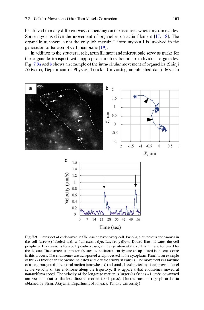
Fig. 7.9a and b shows an example of the intracellular movement of organelles (Shinji
Akiyama, Department of Physics, Tohoku University, unpublished data). Myosin
Fig. 7.9 Transport of endosomes in Chinese hamster ovary cell. Panel a, a numerous endosomes in
the cell (arrows) labeled with a fluorescent dye, Lucifer yellow. Dotted line indicates the cell
periphery. Endosome is formed by endocytosis, an invagination of the cell membrane followed by
the closure. The extracellular materials such as the fluorescent dye are encapsulated in the endosome
in this process. The endosomes are transported and processed in the cytoplasm. Panel b, an example
of the X-Y trace of an endosome indicated with double arrows in Panel a. The movement is a mixture
of a long-range, uni-directional motion (arrowheads) and small, less directed motion (arrows). Panel
c, the velocity of the endosome along the trajectory. It is apparent that endosomes moved at
non-uniform speed. The velocity of the long-rage motion is larger (as fast as ~1 μm/s; downward
arrows) than that of the less directed motion (~0.1 μm/s). (fluorescence micrograph and data
obtained by Shinji Akiyama, Department of Physics, Tohoku University)
7.2 Cellular Movements Other Than Muscle Contraction
105
Some myosins drive the movement of organelles on actin filament [17, 18]. The
organelle transport is not the only job myosin I does: myosin I is involved in the
generation of tension of cell membrane [19].
In addition to the structural role, actin filament and microtubule serve as tracks for
the organelle transport with appropriate motors bound to individual organelles.
Fig. 7.9a and b shows an example of the intracellular movement of organelles (Shinji
Akiyama, Department of Physics, Tohoku University, unpublished data). Myosin
Fig. 7.9 Transport of endosomes in Chinese hamster ovary cell. Panel a, a numerous endosomes in
the cell (arrows) labeled with a fluorescent dye, Lucifer yellow. Dotted line indicates the cell
periphery. Endosome is formed by endocytosis, an invagination of the cell membrane followed by
the closure. The extracellular materials such as the fluorescent dye are encapsulated in the endosome
in this process. The endosomes are transported and processed in the cytoplasm. Panel b, an example
of the X-Y trace of an endosome indicated with double arrows in Panel a. The movement is a mixture
of a long-range, uni-directional motion (arrowheads) and small, less directed motion (arrows). Panel
c, the velocity of the endosome along the trajectory. It is apparent that endosomes moved at
non-uniform speed. The velocity of the long-rage motion is larger (as fast as ~1 μm/s; downward
arrows) than that of the less directed motion (~0.1 μm/s). (fluorescence micrograph and data
obtained by Shinji Akiyama, Department of Physics, Tohoku University)
7.2 Cellular Movements Other Than Muscle Contraction
105