

depolymerization of the polymer. As already described, the monomeric unit of actin
filament is actin monomer (also called G-actin); in the case of microtubules, tubulin
dimers (Sects. 7.3.3 and 7.6.2). Since those filaments are utilized as structural as well
as functional components, the reversible nature of the formation of the filamentous
structure seems to be quite suitable for the cell to respond to the ever-changing
mechanical and/or chemical environment.
The group of intermediate filament system contains several different types of unit
(eg., keratin, vimentin or lamin [10]). The intermediate filaments have been shown to
cooperate with the above two cytoskeletal system [10–12]. Readers are referred to
[13, 14]. Below, we briefly explain non-muscle myosin and from Sect. 7.3., the actin
filament and microtubule system in order to understand their cellular functions in
detail.
7.2.2 Non-muscle Myosins and Organelle Transport
In non-muscle cells, many types of myosin exist. The muscle-type myosin is called
myosin II, as mentioned before, because it was discovered after the first discovery of
a non-muscle myosin (called myosin-I) in Acanthamoeba [15]. Myosin I is singleheaded, has a short, non-alpha helical tail and does not form a filament. Later, other
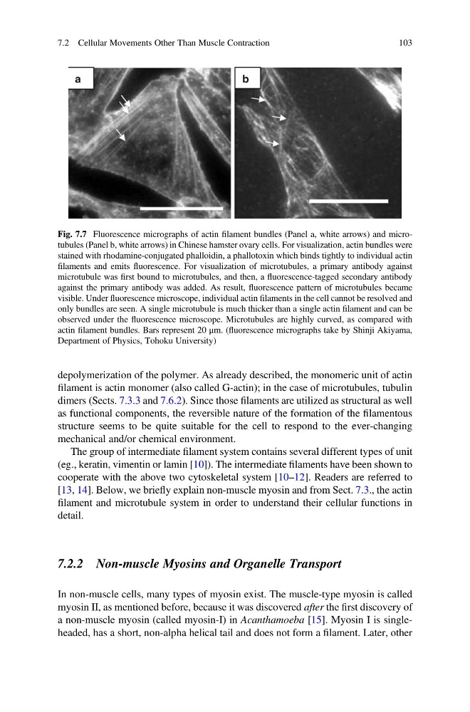
Fig. 7.7 Fluorescence micrographs of actin filament bundles (Panel a, white arrows) and microtubules (Panel b, white arrows) in Chinese hamster ovary cells. For visualization, actin bundles were
stained with rhodamine-conjugated phalloidin, a phallotoxin which binds tightly to individual actin
filaments and emits fluorescence. For visualization of microtubules, a primary antibody against
microtubule was first bound to microtubules, and then, a fluorescence-tagged secondary antibody
against the primary antibody was added. As result, fluorescence pattern of microtubules became
visible. Under fluorescence microscope, individual actin filaments in the cell cannot be resolved and
only bundles are seen. A single microtubule is much thicker than a single actin filament and can be
observed under the fluorescence microscope. Microtubules are highly curved, as compared with
actin filament bundles. Bars represent 20 μm. (fluorescence micrographs take by Shinji Akiyama,
Department of Physics, Tohoku University)
7.2 Cellular Movements Other Than Muscle Contraction
103
filament is actin monomer (also called G-actin); in the case of microtubules, tubulin
dimers (Sects. 7.3.3 and 7.6.2). Since those filaments are utilized as structural as well
as functional components, the reversible nature of the formation of the filamentous
structure seems to be quite suitable for the cell to respond to the ever-changing
mechanical and/or chemical environment.
The group of intermediate filament system contains several different types of unit
(eg., keratin, vimentin or lamin [10]). The intermediate filaments have been shown to
cooperate with the above two cytoskeletal system [10–12]. Readers are referred to
[13, 14]. Below, we briefly explain non-muscle myosin and from Sect. 7.3., the actin
filament and microtubule system in order to understand their cellular functions in
detail.
7.2.2 Non-muscle Myosins and Organelle Transport
In non-muscle cells, many types of myosin exist. The muscle-type myosin is called
myosin II, as mentioned before, because it was discovered after the first discovery of
a non-muscle myosin (called myosin-I) in Acanthamoeba [15]. Myosin I is singleheaded, has a short, non-alpha helical tail and does not form a filament. Later, other
Fig. 7.7 Fluorescence micrographs of actin filament bundles (Panel a, white arrows) and microtubules (Panel b, white arrows) in Chinese hamster ovary cells. For visualization, actin bundles were
stained with rhodamine-conjugated phalloidin, a phallotoxin which binds tightly to individual actin
filaments and emits fluorescence. For visualization of microtubules, a primary antibody against
microtubule was first bound to microtubules, and then, a fluorescence-tagged secondary antibody
against the primary antibody was added. As result, fluorescence pattern of microtubules became
visible. Under fluorescence microscope, individual actin filaments in the cell cannot be resolved and
only bundles are seen. A single microtubule is much thicker than a single actin filament and can be
observed under the fluorescence microscope. Microtubules are highly curved, as compared with
actin filament bundles. Bars represent 20 μm. (fluorescence micrographs take by Shinji Akiyama,
Department of Physics, Tohoku University)
7.2 Cellular Movements Other Than Muscle Contraction
103