

Heavy
chains
Head
Myosin II
Myosin II associated
by the tail region
Myosin II filament
Light chains (total four)
Head (globular domain)
Tail (coiled coil α-helix)
b
a
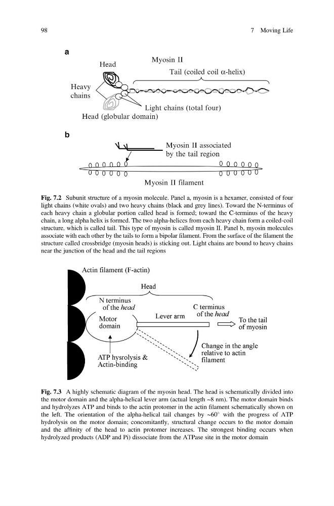
Fig. 7.2 Subunit structure of a myosin molecule. Panel a, myosin is a hexamer, consisted of four
light chains (white ovals) and two heavy chains (black and grey lines). Toward the N-terminus of
each heavy chain a globular portion called head is formed; toward the C-terminus of the heavy
chain, a long alpha helix is formed. The two alpha-helices from each heavy chain form a coiled-coil
structure, which is called tail. This type of myosin is called myosin II. Panel b, myosin molecules
associate with each other by the tails to form a bipolar filament. From the surface of the filament the
structure called crossbridge (myosin heads) is sticking out. Light chains are bound to heavy chains
near the junction of the head and the tail regions
Fig. 7.3 A highly schematic diagram of the myosin head. The head is schematically divided into
the motor domain and the alpha-helical lever arm (actual length ~8 nm). The motor domain binds
and hydrolyzes ATP and binds to the actin protomer in the actin filament schematically shown on
the left. The orientation of the alpha-helical tail changes by ~60
with the progress of ATP
hydrolysis on the motor domain; concomitantly, structural change occurs to the motor domain
and the affinity of the head to actin protomer increases. The strongest binding occurs when
hydrolyzed products (ADP and Pi) dissociate from the ATPase site in the motor domain
98
7 Moving Life
chains
Head
Myosin II
Myosin II associated
by the tail region
Myosin II filament
Light chains (total four)
Head (globular domain)
Tail (coiled coil α-helix)
b
a
Fig. 7.2 Subunit structure of a myosin molecule. Panel a, myosin is a hexamer, consisted of four
light chains (white ovals) and two heavy chains (black and grey lines). Toward the N-terminus of
each heavy chain a globular portion called head is formed; toward the C-terminus of the heavy
chain, a long alpha helix is formed. The two alpha-helices from each heavy chain form a coiled-coil
structure, which is called tail. This type of myosin is called myosin II. Panel b, myosin molecules
associate with each other by the tails to form a bipolar filament. From the surface of the filament the
structure called crossbridge (myosin heads) is sticking out. Light chains are bound to heavy chains
near the junction of the head and the tail regions
Fig. 7.3 A highly schematic diagram of the myosin head. The head is schematically divided into
the motor domain and the alpha-helical lever arm (actual length ~8 nm). The motor domain binds
and hydrolyzes ATP and binds to the actin protomer in the actin filament schematically shown on
the left. The orientation of the alpha-helical tail changes by ~60
with the progress of ATP
hydrolysis on the motor domain; concomitantly, structural change occurs to the motor domain
and the affinity of the head to actin protomer increases. The strongest binding occurs when
hydrolyzed products (ADP and Pi) dissociate from the ATPase site in the motor domain
98
7 Moving Life