

Before
contraction
Thick (myosin) filament
I A
M
Z
Sarcomere
Thin (actin) filament
e
d
c
b
a
After
contraction
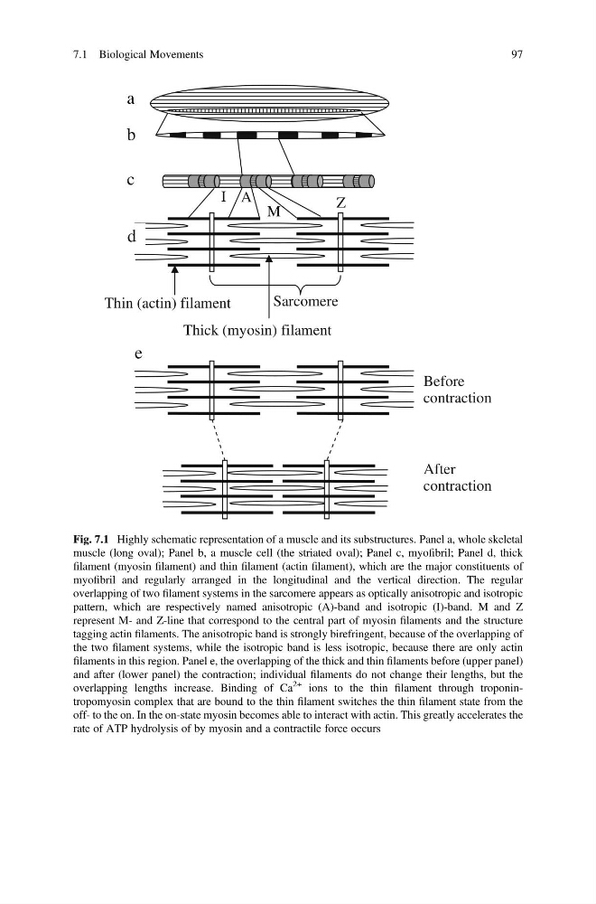
Fig. 7.1 Highly schematic representation of a muscle and its substructures. Panel a, whole skeletal
muscle (long oval); Panel b, a muscle cell (the striated oval); Panel c, myofibril; Panel d, thick
filament (myosin filament) and thin filament (actin filament), which are the major constituents of
myofibril and regularly arranged in the longitudinal and the vertical direction. The regular
overlapping of two filament systems in the sarcomere appears as optically anisotropic and isotropic
pattern, which are respectively named anisotropic (A)-band and isotropic (I)-band. M and Z
represent M- and Z-line that correspond to the central part of myosin filaments and the structure
tagging actin filaments. The anisotropic band is strongly birefringent, because of the overlapping of
the two filament systems, while the isotropic band is less isotropic, because there are only actin
filaments in this region. Panel e, the overlapping of the thick and thin filaments before (upper panel)
and after (lower panel) the contraction; individual filaments do not change their lengths, but the
overlapping lengths increase. Binding of Ca
2+ ions to the thin filament through troponintropomyosin complex that are bound to the thin filament switches the thin filament state from the
off- to the on. In the on-state myosin becomes able to interact with actin. This greatly accelerates the
rate of ATP hydrolysis of by myosin and a contractile force occurs
7.1 Biological Movements
97
contraction
Thick (myosin) filament
I A
M
Z
Sarcomere
Thin (actin) filament
e
d
c
b
a
After
contraction
Fig. 7.1 Highly schematic representation of a muscle and its substructures. Panel a, whole skeletal
muscle (long oval); Panel b, a muscle cell (the striated oval); Panel c, myofibril; Panel d, thick
filament (myosin filament) and thin filament (actin filament), which are the major constituents of
myofibril and regularly arranged in the longitudinal and the vertical direction. The regular
overlapping of two filament systems in the sarcomere appears as optically anisotropic and isotropic
pattern, which are respectively named anisotropic (A)-band and isotropic (I)-band. M and Z
represent M- and Z-line that correspond to the central part of myosin filaments and the structure
tagging actin filaments. The anisotropic band is strongly birefringent, because of the overlapping of
the two filament systems, while the isotropic band is less isotropic, because there are only actin
filaments in this region. Panel e, the overlapping of the thick and thin filaments before (upper panel)
and after (lower panel) the contraction; individual filaments do not change their lengths, but the
overlapping lengths increase. Binding of Ca
2+ ions to the thin filament through troponintropomyosin complex that are bound to the thin filament switches the thin filament state from the
off- to the on. In the on-state myosin becomes able to interact with actin. This greatly accelerates the
rate of ATP hydrolysis of by myosin and a contractile force occurs
7.1 Biological Movements
97