

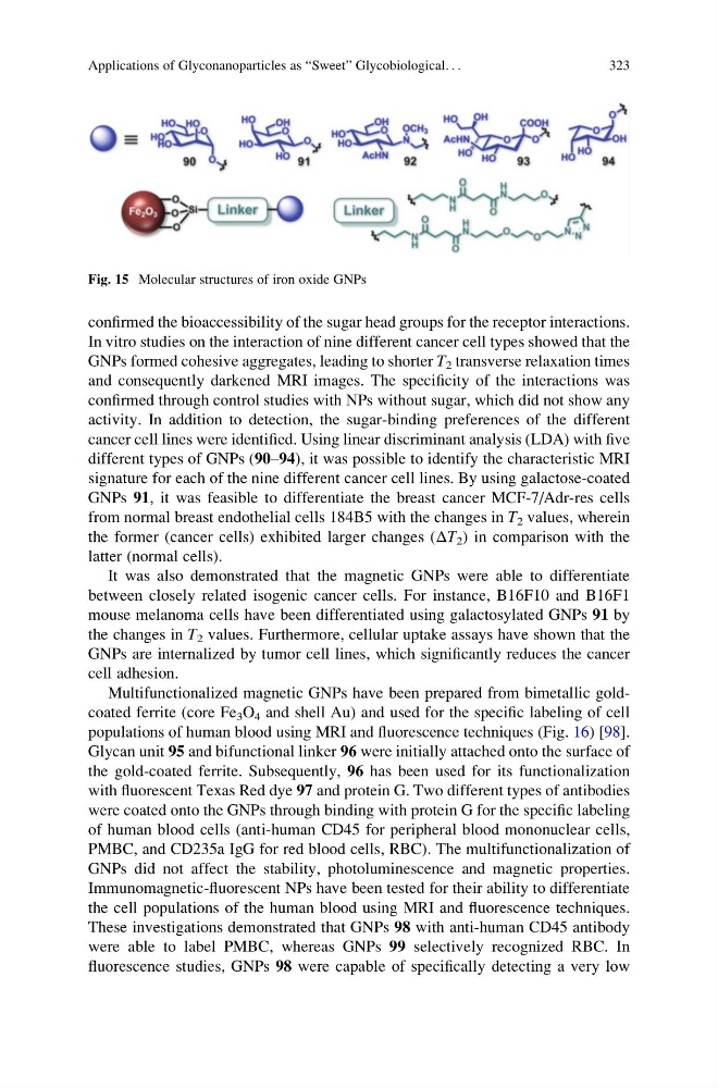
confirmed the bioaccessibility of the sugar head groups for the receptor interactions.
In vitro studies on the interaction of nine different cancer cell types showed that the
GNPs formed cohesive aggregates, leading to shorter T 2 transverse relaxation times
and consequently darkened MRI images. The specificity of the interactions was
confirmed through control studies with NPs without sugar, which did not show any
activity. In addition to detection, the sugar-binding preferences of the different
cancer cell lines were identified. Using linear discriminant analysis (LDA) with five
different types of GNPs (90–94), it was possible to identify the characteristic MRI
signature for each of the nine different cancer cell lines. By using galactose-coated
GNPs 91, it was feasible to differentiate the breast cancer MCF-7/Adr-res cells
from normal breast endothelial cells 184B5 with the changes in T 2 values, wherein
the former (cancer cells) exhibited larger changes (ΔT 2 ) in comparison with the
latter (normal cells).
It was also demonstrated that the magnetic GNPs were able to differentiate
between closely related isogenic cancer cells. For instance, B16F10 and B16F1
mouse melanoma cells have been differentiated using galactosylated GNPs 91 by
the changes in T 2 values. Furthermore, cellular uptake assays have shown that the
GNPs are internalized by tumor cell lines, which significantly reduces the cancer
cell adhesion.
Multifunctionalized magnetic GNPs have been prepared from bimetallic goldcoated ferrite (core Fe 3 O 4 and shell Au) and used for the specific labeling of cell
populations of human blood using MRI and fluorescence techniques (Fig. 16) [98].
Glycan unit 95 and bifunctional linker 96 were initially attached onto the surface of
the gold-coated ferrite. Subsequently, 96 has been used for its functionalization
with fluorescent Texas Red dye 97 and protein G. Two different types of antibodies
were coated onto the GNPs through binding with protein G for the specific labeling
of human blood cells (anti-human CD45 for peripheral blood mononuclear cells,
PMBC, and CD235a IgG for red blood cells, RBC). The multifunctionalization of
GNPs did not affect the stability, photoluminescence and magnetic properties.
Immunomagnetic-fluorescent NPs have been tested for their ability to differentiate
the cell populations of the human blood using MRI and fluorescence techniques.
These investigations demonstrated that GNPs 98 with anti-human CD45 antibody
were able to label PMBC, whereas GNPs 99 selectively recognized RBC. In
fluorescence studies, GNPs 98 were capable of specifically detecting a very low
Fig. 15 Molecular structures of iron oxide GNPs
Applications of Glyconanoparticles as “Sweet” Glycobiological. . .
323
In vitro studies on the interaction of nine different cancer cell types showed that the
GNPs formed cohesive aggregates, leading to shorter T 2 transverse relaxation times
and consequently darkened MRI images. The specificity of the interactions was
confirmed through control studies with NPs without sugar, which did not show any
activity. In addition to detection, the sugar-binding preferences of the different
cancer cell lines were identified. Using linear discriminant analysis (LDA) with five
different types of GNPs (90–94), it was possible to identify the characteristic MRI
signature for each of the nine different cancer cell lines. By using galactose-coated
GNPs 91, it was feasible to differentiate the breast cancer MCF-7/Adr-res cells
from normal breast endothelial cells 184B5 with the changes in T 2 values, wherein
the former (cancer cells) exhibited larger changes (ΔT 2 ) in comparison with the
latter (normal cells).
It was also demonstrated that the magnetic GNPs were able to differentiate
between closely related isogenic cancer cells. For instance, B16F10 and B16F1
mouse melanoma cells have been differentiated using galactosylated GNPs 91 by
the changes in T 2 values. Furthermore, cellular uptake assays have shown that the
GNPs are internalized by tumor cell lines, which significantly reduces the cancer
cell adhesion.
Multifunctionalized magnetic GNPs have been prepared from bimetallic goldcoated ferrite (core Fe 3 O 4 and shell Au) and used for the specific labeling of cell
populations of human blood using MRI and fluorescence techniques (Fig. 16) [98].
Glycan unit 95 and bifunctional linker 96 were initially attached onto the surface of
the gold-coated ferrite. Subsequently, 96 has been used for its functionalization
with fluorescent Texas Red dye 97 and protein G. Two different types of antibodies
were coated onto the GNPs through binding with protein G for the specific labeling
of human blood cells (anti-human CD45 for peripheral blood mononuclear cells,
PMBC, and CD235a IgG for red blood cells, RBC). The multifunctionalization of
GNPs did not affect the stability, photoluminescence and magnetic properties.
Immunomagnetic-fluorescent NPs have been tested for their ability to differentiate
the cell populations of the human blood using MRI and fluorescence techniques.
These investigations demonstrated that GNPs 98 with anti-human CD45 antibody
were able to label PMBC, whereas GNPs 99 selectively recognized RBC. In
fluorescence studies, GNPs 98 were capable of specifically detecting a very low
Fig. 15 Molecular structures of iron oxide GNPs
Applications of Glyconanoparticles as “Sweet” Glycobiological. . .
323