

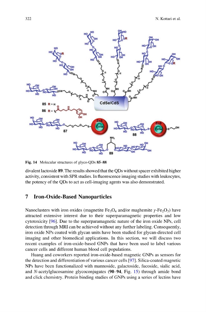
divalent lactoside 89. The results showed that the QDs without spacer exhibited higher
activity, consistent with SPR studies. In fluorescence imaging studies with leukocytes,
the potency of the QDs to act as cell-imaging agents was also demonstrated.
7 Iron-Oxide-Based Nanoparticles
Nanoclusters with iron oxides (magnetite Fe 3 O 4 and/or maghemite γ-Fe 2 O 3 ) have
attracted extensive interest due to their superparamagnetic properties and low
cytotoxicity [96]. Due to the superparamagnetic nature of the iron oxide NPs, cell
detection through MRI can be achieved without any further labeling. Consequently,
iron oxide NPs coated with glycan units have been studied for glycan-directed cell
imaging and other biomedical applications. In this section, we will discuss two
recent examples of iron-oxide-based GNPs that have been used to label various
cancer cells and different human blood cell populations.
Huang and coworkers reported iron-oxide-based magnetic GNPs as sensors for
the detection and differentiation of various cancer cells [97]. Silica-coated magnetic
NPs have been functionalized with mannoside, galactoside, fucoside, sialic acid,
and N-acetylglucosamine glycoconjugates (90–94, Fig. 15) through amide bond
and click chemistry. Protein binding studies of GNPs using a series of lectins have
Fig. 14 Molecular structures of glyco-QDs 85–88
322
N. Kottari et al.
activity, consistent with SPR studies. In fluorescence imaging studies with leukocytes,
the potency of the QDs to act as cell-imaging agents was also demonstrated.
7 Iron-Oxide-Based Nanoparticles
Nanoclusters with iron oxides (magnetite Fe 3 O 4 and/or maghemite γ-Fe 2 O 3 ) have
attracted extensive interest due to their superparamagnetic properties and low
cytotoxicity [96]. Due to the superparamagnetic nature of the iron oxide NPs, cell
detection through MRI can be achieved without any further labeling. Consequently,
iron oxide NPs coated with glycan units have been studied for glycan-directed cell
imaging and other biomedical applications. In this section, we will discuss two
recent examples of iron-oxide-based GNPs that have been used to label various
cancer cells and different human blood cell populations.
Huang and coworkers reported iron-oxide-based magnetic GNPs as sensors for
the detection and differentiation of various cancer cells [97]. Silica-coated magnetic
NPs have been functionalized with mannoside, galactoside, fucoside, sialic acid,
and N-acetylglucosamine glycoconjugates (90–94, Fig. 15) through amide bond
and click chemistry. Protein binding studies of GNPs using a series of lectins have
Fig. 14 Molecular structures of glyco-QDs 85–88
322
N. Kottari et al.