

mechanism for the fluorescence quenching was unclear, the dissociation constants of
QRs 85 derived from the fluorescence quenching studies was in the order of 10
7
,
which was three to four orders of magnitude higher than that of the β-D-galactoside
monomers, indicating the enhanced interactions of the clustered QRs with lectin. The
specificity of the interactions was confirmed by competitive studies through the
addition of galactose, which caused complete recovery of the fluorescence intensity
of the QRs. In vitro enzyme hydrolysis studies using porcine liver esterase, the
regulated release of dopamine, demonstrated the potency of QRs 86 to act as drug
delivery agents. In biological studies, glyco-QRs have been selectively recognized by
human nasopharyngeal epidermal carcinoma (KB) cells through the GLUT-1 transporter, a glucose receptor found at the outer KB cell membrane. In the assays, cellular
uptake of the QRs was confirmed from their high internalization values. The
specificities of glyco-QRs in in vivo assays have been confirmed through competitive
studies with the free galactose.
QDs functionalized with lactose have been studied for protein interactions, cell
imaging, and anti adhesive applications [95]. Lactose-QDs have been prepared with
and without PEG spacer (87 and 88, respectively). In the case of 88 without spacer,
QDs with different lactoside densities were synthesized in combination with
ethanethiol (Fig. 14). The amount of sugar loading was determined from NMR studies
in conjunction with thermogravimetric analysis (TGA). The protein binding studies of
QDs were performed in SPR assays over the surface of the immobilized PNA lectin.
Glyco-QDs showed higher affinity than free lactose and, among the glyco-QDs, the
affinity was lower for the QDs 87 having PEG spacer as compared to 88 deprived of
spacer, thus demonstrating the importance of linker design in multivalent
glycoconjugates. The anti-adhesive ability of glyco-QDs to prevent the adhesion of
human acute monocytic leukemia cells to human umbilical vein endothelial cells
(HUVEC) was measured. These cells play a critical role in the pathogenesis of septic
shock. In the assays, glyco-QDs exhibited five to sixfold higher activity than the
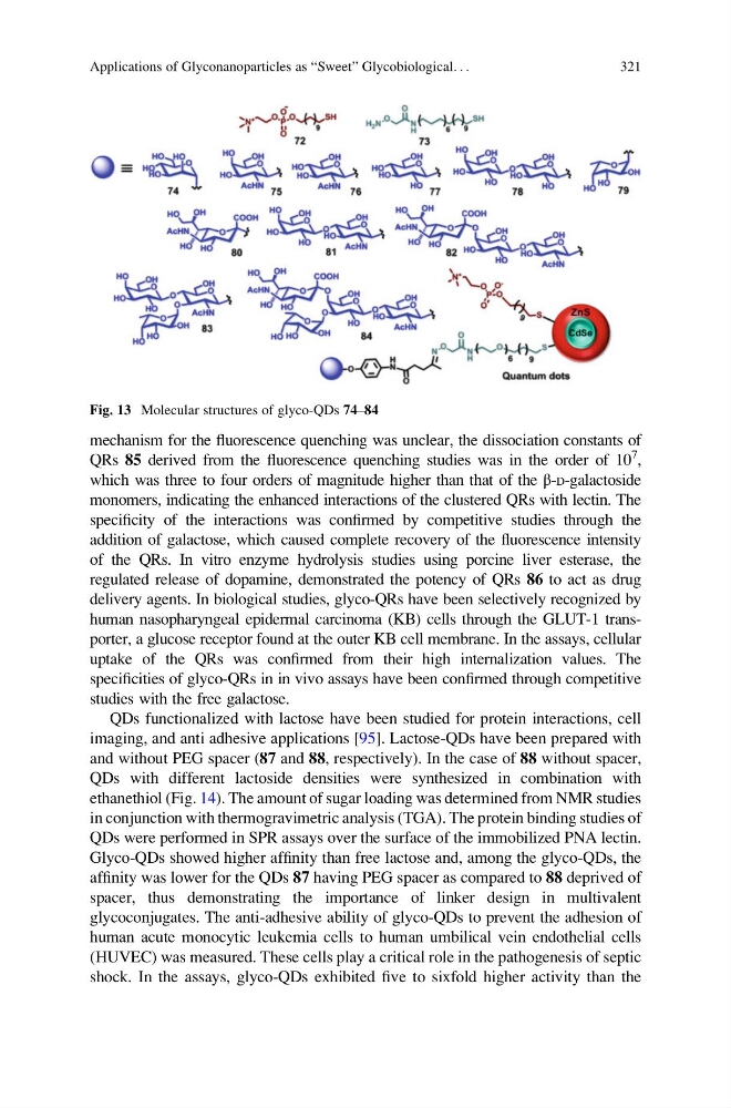
Fig. 13 Molecular structures of glyco-QDs 74–84
Applications of Glyconanoparticles as “Sweet” Glycobiological. . .
321
QRs 85 derived from the fluorescence quenching studies was in the order of 10
7
,
which was three to four orders of magnitude higher than that of the β-D-galactoside
monomers, indicating the enhanced interactions of the clustered QRs with lectin. The
specificity of the interactions was confirmed by competitive studies through the
addition of galactose, which caused complete recovery of the fluorescence intensity
of the QRs. In vitro enzyme hydrolysis studies using porcine liver esterase, the
regulated release of dopamine, demonstrated the potency of QRs 86 to act as drug
delivery agents. In biological studies, glyco-QRs have been selectively recognized by
human nasopharyngeal epidermal carcinoma (KB) cells through the GLUT-1 transporter, a glucose receptor found at the outer KB cell membrane. In the assays, cellular
uptake of the QRs was confirmed from their high internalization values. The
specificities of glyco-QRs in in vivo assays have been confirmed through competitive
studies with the free galactose.
QDs functionalized with lactose have been studied for protein interactions, cell
imaging, and anti adhesive applications [95]. Lactose-QDs have been prepared with
and without PEG spacer (87 and 88, respectively). In the case of 88 without spacer,
QDs with different lactoside densities were synthesized in combination with
ethanethiol (Fig. 14). The amount of sugar loading was determined from NMR studies
in conjunction with thermogravimetric analysis (TGA). The protein binding studies of
QDs were performed in SPR assays over the surface of the immobilized PNA lectin.
Glyco-QDs showed higher affinity than free lactose and, among the glyco-QDs, the
affinity was lower for the QDs 87 having PEG spacer as compared to 88 deprived of
spacer, thus demonstrating the importance of linker design in multivalent
glycoconjugates. The anti-adhesive ability of glyco-QDs to prevent the adhesion of
human acute monocytic leukemia cells to human umbilical vein endothelial cells
(HUVEC) was measured. These cells play a critical role in the pathogenesis of septic
shock. In the assays, glyco-QDs exhibited five to sixfold higher activity than the
Fig. 13 Molecular structures of glyco-QDs 74–84
Applications of Glyconanoparticles as “Sweet” Glycobiological. . .
321