

hybrid materials with modular organic composition and functionality. Several
methods have been developed to modify the magnetite nanoparticles. The most
commonly used method is to encapsulate magnetic nanoparticles in a polymeric
matrix, such as polylactic acid (PLA) or polylactic-co-glycolic acid (PLGA)
microspheres [98], Therefore, many approaches have been focused on the encapsulation of magnetic nanoparticles with biocompatible materials such as dextran,
chitosan, gelatin, poly(ethylene glycol) (PEG), PLGA, poly(D,L-lactide) (PLA),
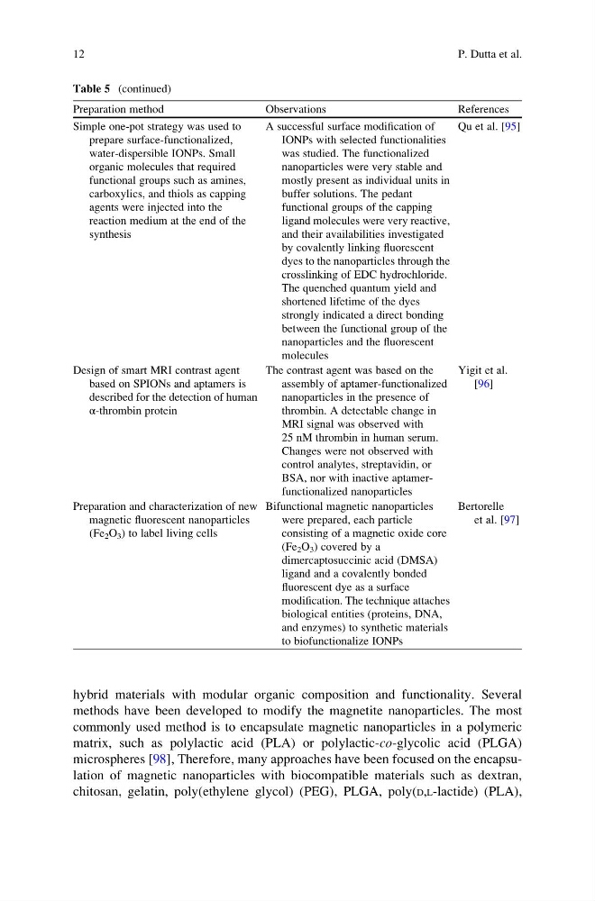
Table 5 (continued)
Preparation method
Observations
References
Simple one-pot strategy was used to
prepare surface-functionalized,
water-dispersible IONPs. Small
organic molecules that required
functional groups such as amines,
carboxylics, and thiols as capping
agents were injected into the
reaction medium at the end of the
synthesis
A successful surface modification of
IONPs with selected functionalities
was studied. The functionalized
nanoparticles were very stable and
mostly present as individual units in
buffer solutions. The pedant
functional groups of the capping
ligand molecules were very reactive,
and their availabilities investigated
by covalently linking fluorescent
dyes to the nanoparticles through the
crosslinking of EDC hydrochloride.
The quenched quantum yield and
shortened lifetime of the dyes
strongly indicated a direct bonding
between the functional group of the
nanoparticles and the fluorescent
molecules
Qu et al. [95]
Design of smart MRI contrast agent
based on SPIONs and aptamers is
described for the detection of human
α-thrombin protein
The contrast agent was based on the
assembly of aptamer-functionalized
nanoparticles in the presence of
thrombin. A detectable change in
MRI signal was observed with
25 nM thrombin in human serum.
Changes were not observed with
control analytes, streptavidin, or
BSA, nor with inactive aptamerfunctionalized nanoparticles
Yigit et al.
[96]
Preparation and characterization of new
magnetic fluorescent nanoparticles
(Fe 2 O 3 ) to label living cells
Bifunctional magnetic nanoparticles
were prepared, each particle
consisting of a magnetic oxide core
(Fe 2 O 3 ) covered by a
dimercaptosuccinic acid (DMSA)
ligand and a covalently bonded
fluorescent dye as a surface
modification. The technique attaches
biological entities (proteins, DNA,
and enzymes) to synthetic materials
to biofunctionalize IONPs
Bertorelle
et al. [97]
12
P. Dutta et al.
methods have been developed to modify the magnetite nanoparticles. The most
commonly used method is to encapsulate magnetic nanoparticles in a polymeric
matrix, such as polylactic acid (PLA) or polylactic-co-glycolic acid (PLGA)
microspheres [98], Therefore, many approaches have been focused on the encapsulation of magnetic nanoparticles with biocompatible materials such as dextran,
chitosan, gelatin, poly(ethylene glycol) (PEG), PLGA, poly(D,L-lactide) (PLA),
Table 5 (continued)
Preparation method
Observations
References
Simple one-pot strategy was used to
prepare surface-functionalized,
water-dispersible IONPs. Small
organic molecules that required
functional groups such as amines,
carboxylics, and thiols as capping
agents were injected into the
reaction medium at the end of the
synthesis
A successful surface modification of
IONPs with selected functionalities
was studied. The functionalized
nanoparticles were very stable and
mostly present as individual units in
buffer solutions. The pedant
functional groups of the capping
ligand molecules were very reactive,
and their availabilities investigated
by covalently linking fluorescent
dyes to the nanoparticles through the
crosslinking of EDC hydrochloride.
The quenched quantum yield and
shortened lifetime of the dyes
strongly indicated a direct bonding
between the functional group of the
nanoparticles and the fluorescent
molecules
Qu et al. [95]
Design of smart MRI contrast agent
based on SPIONs and aptamers is
described for the detection of human
α-thrombin protein
The contrast agent was based on the
assembly of aptamer-functionalized
nanoparticles in the presence of
thrombin. A detectable change in
MRI signal was observed with
25 nM thrombin in human serum.
Changes were not observed with
control analytes, streptavidin, or
BSA, nor with inactive aptamerfunctionalized nanoparticles
Yigit et al.
[96]
Preparation and characterization of new
magnetic fluorescent nanoparticles
(Fe 2 O 3 ) to label living cells
Bifunctional magnetic nanoparticles
were prepared, each particle
consisting of a magnetic oxide core
(Fe 2 O 3 ) covered by a
dimercaptosuccinic acid (DMSA)
ligand and a covalently bonded
fluorescent dye as a surface
modification. The technique attaches
biological entities (proteins, DNA,
and enzymes) to synthetic materials
to biofunctionalize IONPs
Bertorelle
et al. [97]
12
P. Dutta et al.