

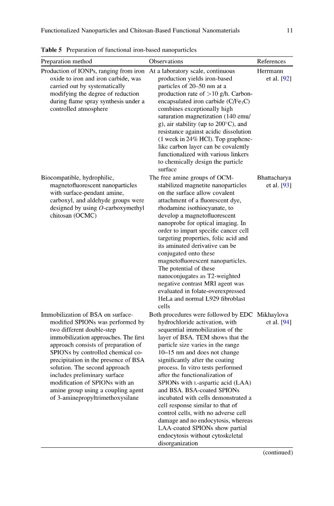
Table 5 Preparation of functional iron-based nanoparticles
Preparation method
Observations
References
Production of IONPs, ranging from iron
oxide to iron and iron carbide, was
carried out by systematically
modifying the degree of reduction
during flame spray synthesis under a
controlled atmosphere
At a laboratory scale, continuous
production yields iron-based
particles of 20–50 nm at a
production rate of >10 g/h. Carbonencapsulated iron carbide (C/Fe 3 C)
combines exceptionally high
saturation magnetization (140 emu/
g), air stability (up to 200
C), and
resistance against acidic dissolution
(1 week in 24% HCl). Top graphenelike carbon layer can be covalently
functionalized with various linkers
to chemically design the particle
surface
Herrmann
et al. [92]
Biocompatible, hydrophilic,
magnetofluorescent nanoparticles
with surface-pendant amine,
carboxyl, and aldehyde groups were
designed by using O-carboxymethyl
chitosan (OCMC)
The free amine groups of OCMstabilized magnetite nanoparticles
on the surface allow covalent
attachment of a fluorescent dye,
rhodamine isothiocyanate, to
develop a magnetofluorescent
nanoprobe for optical imaging. In
order to impart specific cancer cell
targeting properties, folic acid and
its aminated derivative can be
conjugated onto these
magnetofluorescent nanoparticles.
The potential of these
nanoconjugates as T2-weighted
negative contrast MRI agent was
evaluated in folate-overexpressed
HeLa and normal L929 fibroblast
cells
Bhattacharya
et al. [93]
Immobilization of BSA on surfacemodified SPIONs was performed by
two different double-step
immobilization approaches. The first
approach consists of preparation of
SPIONs by controlled chemical coprecipitation in the presence of BSA
solution. The second approach
includes preliminary surface
modification of SPIONs with an
amine group using a coupling agent
of 3-aminepropyltrimethoxysilane
Both procedures were followed by EDC
hydrochloride activation, with
sequential immobilization of the
layer of BSA. TEM shows that the
particle size varies in the range
10–15 nm and does not change
significantly after the coating
process. In vitro tests performed
after the functionalization of
SPIONs with L-aspartic acid (LAA)
and BSA. BSA-coated SPIONs
incubated with cells demonstrated a
cell response similar to that of
control cells, with no adverse cell
damage and no endocytosis, whereas
LAA-coated SPIONs show partial
endocytosis without cytoskeletal
disorganization
Mikhaylova
et al. [94]
(continued)
Functionalized Nanoparticles and Chitosan-Based Functional Nanomaterials
11
Preparation method
Observations
References
Production of IONPs, ranging from iron
oxide to iron and iron carbide, was
carried out by systematically
modifying the degree of reduction
during flame spray synthesis under a
controlled atmosphere
At a laboratory scale, continuous
production yields iron-based
particles of 20–50 nm at a
production rate of >10 g/h. Carbonencapsulated iron carbide (C/Fe 3 C)
combines exceptionally high
saturation magnetization (140 emu/
g), air stability (up to 200
C), and
resistance against acidic dissolution
(1 week in 24% HCl). Top graphenelike carbon layer can be covalently
functionalized with various linkers
to chemically design the particle
surface
Herrmann
et al. [92]
Biocompatible, hydrophilic,
magnetofluorescent nanoparticles
with surface-pendant amine,
carboxyl, and aldehyde groups were
designed by using O-carboxymethyl
chitosan (OCMC)
The free amine groups of OCMstabilized magnetite nanoparticles
on the surface allow covalent
attachment of a fluorescent dye,
rhodamine isothiocyanate, to
develop a magnetofluorescent
nanoprobe for optical imaging. In
order to impart specific cancer cell
targeting properties, folic acid and
its aminated derivative can be
conjugated onto these
magnetofluorescent nanoparticles.
The potential of these
nanoconjugates as T2-weighted
negative contrast MRI agent was
evaluated in folate-overexpressed
HeLa and normal L929 fibroblast
cells
Bhattacharya
et al. [93]
Immobilization of BSA on surfacemodified SPIONs was performed by
two different double-step
immobilization approaches. The first
approach consists of preparation of
SPIONs by controlled chemical coprecipitation in the presence of BSA
solution. The second approach
includes preliminary surface
modification of SPIONs with an
amine group using a coupling agent
of 3-aminepropyltrimethoxysilane
Both procedures were followed by EDC
hydrochloride activation, with
sequential immobilization of the
layer of BSA. TEM shows that the
particle size varies in the range
10–15 nm and does not change
significantly after the coating
process. In vitro tests performed
after the functionalization of
SPIONs with L-aspartic acid (LAA)
and BSA. BSA-coated SPIONs
incubated with cells demonstrated a
cell response similar to that of
control cells, with no adverse cell
damage and no endocytosis, whereas
LAA-coated SPIONs show partial
endocytosis without cytoskeletal
disorganization
Mikhaylova
et al. [94]
(continued)
Functionalized Nanoparticles and Chitosan-Based Functional Nanomaterials
11