

cross the M cells in the Peyer’s patch and the mesentery on the surface of the
gastrointestinal mucosa for delivering the drug to the systemic circulation [34].
Accordingly, particles larger than 200 nm can be mechanically filtered in the spleen
whereas those smaller than 100 nm leave the blood vessels through fenestrations in
the endothelial lining. Therefore, nanoparticles with relatively small size (in the
range of 100–200 nm) are desirable for delivery of drugs to tumors [35, 36].
Nanoparticles can be prepared by the same methods as those described for
microparticles, but by using a comparatively small ratio of dispersed phase:
0
1 0
2 0
3 0
4 0
5 0
0
20
40
60
80
100
t / h
PCL
PCL-30B
PCL-C18
PCL-6h
PCL-C18-0h
PCL-C18-6h
PCL-30B-0h
PCL-30B-6h
PCL-0h
a
b
Wt. Loss / %
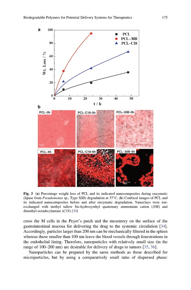
Fig. 3 (a) Percentage weight loss of PCL and its indicated nanocomposites during enzymatic
(lipase from Pseudomonas sp., Type XIII) degradation at 37
C. (b) Confocal images of PCL and
its indicated nanocomposites before and after enzymatic degradation. Nanoclays were ionexchanged with methyl tallow bis-hydroxyethyl quaternary ammonium cation (30B) and
dimethyl-octadecylamine (C18) [30]
Biodegradable Polymers for Potential Delivery Systems for Therapeutics
175
gastrointestinal mucosa for delivering the drug to the systemic circulation [34].
Accordingly, particles larger than 200 nm can be mechanically filtered in the spleen
whereas those smaller than 100 nm leave the blood vessels through fenestrations in
the endothelial lining. Therefore, nanoparticles with relatively small size (in the
range of 100–200 nm) are desirable for delivery of drugs to tumors [35, 36].
Nanoparticles can be prepared by the same methods as those described for
microparticles, but by using a comparatively small ratio of dispersed phase:
0
1 0
2 0
3 0
4 0
5 0
0
20
40
60
80
100
t / h
PCL
PCL-30B
PCL-C18
PCL-6h
PCL-C18-0h
PCL-C18-6h
PCL-30B-0h
PCL-30B-6h
PCL-0h
a
b
Wt. Loss / %
Fig. 3 (a) Percentage weight loss of PCL and its indicated nanocomposites during enzymatic
(lipase from Pseudomonas sp., Type XIII) degradation at 37
C. (b) Confocal images of PCL and
its indicated nanocomposites before and after enzymatic degradation. Nanoclays were ionexchanged with methyl tallow bis-hydroxyethyl quaternary ammonium cation (30B) and
dimethyl-octadecylamine (C18) [30]
Biodegradable Polymers for Potential Delivery Systems for Therapeutics
175