

assembly line [16]. It consists of three independent PX–JX 2 devices that have been
incorporated into a large programmed 2D DNA surface, known as DNA origami
[17]. DNA origami consists of a long scaffold strand, typically M13 single-stranded
DNA (about 7,500 nucleotides) and about 200 “staple strands” that fold it into its
shape; each can be addressed individually. Thus, specific PX–JX 2 devices are
located in each of the three positions near the top of the origami construct, and
each carries a specific cargo. The three cargoes (left to right in Fig. 10) are a 5 nm
gold nanoparticle, a pair of coupled 5 nm gold nanoparticles, and a 10 nm gold
nanoparticle. The bottom of the construct contains a row of sites where a
somersaulting tensegrity triangle walker can pass by the three cargo stations. If
the PX–JX 2 device is in the JX 2 conformation, nothing will happen when the walker
passes the cargo station. If the walker is in the PX conformation, the cargo will be
transferred to the walker. Thus, depending on the programming of the assembly
line, eight different products (2
3 ) can be produced. Figure 10 shows the assembly of
the product consisting of adding all cargoes to the walker. The right-hand column
shows AFM images of the assembly line that correspond to the schematics in the
middle column.
I have tried to give a flavor of structural DNA nanotechnology by illustrations of
work from my own laboratory. A decade ago this would have been a very complete
coverage of this field. However, the field has grown enormously since then, and
much more has been done. There are numerous laboratories worldwide that have
participated in this enterprise, and each contains many workers, not just a principal
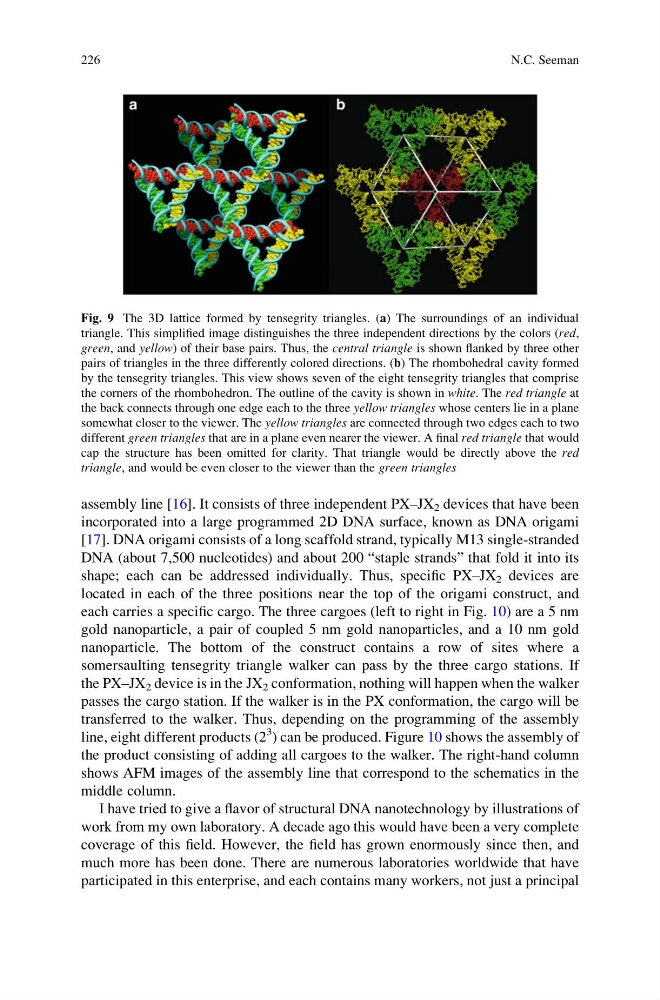
Fig. 9 The 3D lattice formed by tensegrity triangles. (a) The surroundings of an individual
triangle. This simplified image distinguishes the three independent directions by the colors (red,
green, and yellow) of their base pairs. Thus, the central triangle is shown flanked by three other
pairs of triangles in the three differently colored directions. (b) The rhombohedral cavity formed
by the tensegrity triangles. This view shows seven of the eight tensegrity triangles that comprise
the corners of the rhombohedron. The outline of the cavity is shown in white. The red triangle at
the back connects through one edge each to the three yellow triangles whose centers lie in a plane
somewhat closer to the viewer. The yellow triangles are connected through two edges each to two
different green triangles that are in a plane even nearer the viewer. A final red triangle that would
cap the structure has been omitted for clarity. That triangle would be directly above the red
triangle, and would be even closer to the viewer than the green triangles
226
N.C. Seeman
incorporated into a large programmed 2D DNA surface, known as DNA origami
[17]. DNA origami consists of a long scaffold strand, typically M13 single-stranded
DNA (about 7,500 nucleotides) and about 200 “staple strands” that fold it into its
shape; each can be addressed individually. Thus, specific PX–JX 2 devices are
located in each of the three positions near the top of the origami construct, and
each carries a specific cargo. The three cargoes (left to right in Fig. 10) are a 5 nm
gold nanoparticle, a pair of coupled 5 nm gold nanoparticles, and a 10 nm gold
nanoparticle. The bottom of the construct contains a row of sites where a
somersaulting tensegrity triangle walker can pass by the three cargo stations. If
the PX–JX 2 device is in the JX 2 conformation, nothing will happen when the walker
passes the cargo station. If the walker is in the PX conformation, the cargo will be
transferred to the walker. Thus, depending on the programming of the assembly
line, eight different products (2
3 ) can be produced. Figure 10 shows the assembly of
the product consisting of adding all cargoes to the walker. The right-hand column
shows AFM images of the assembly line that correspond to the schematics in the
middle column.
I have tried to give a flavor of structural DNA nanotechnology by illustrations of
work from my own laboratory. A decade ago this would have been a very complete
coverage of this field. However, the field has grown enormously since then, and
much more has been done. There are numerous laboratories worldwide that have
participated in this enterprise, and each contains many workers, not just a principal
Fig. 9 The 3D lattice formed by tensegrity triangles. (a) The surroundings of an individual
triangle. This simplified image distinguishes the three independent directions by the colors (red,
green, and yellow) of their base pairs. Thus, the central triangle is shown flanked by three other
pairs of triangles in the three differently colored directions. (b) The rhombohedral cavity formed
by the tensegrity triangles. This view shows seven of the eight tensegrity triangles that comprise
the corners of the rhombohedron. The outline of the cavity is shown in white. The red triangle at
the back connects through one edge each to the three yellow triangles whose centers lie in a plane
somewhat closer to the viewer. The yellow triangles are connected through two edges each to two
different green triangles that are in a plane even nearer the viewer. A final red triangle that would
cap the structure has been omitted for clarity. That triangle would be directly above the red
triangle, and would be even closer to the viewer than the green triangles
226
N.C. Seeman