

of three types of subunits FN-I, FN-II, and FN-III that differ in the number of amino
acids (45, 60, and 90, respectively) [127]. Fibronectin contains 220 kDa subunits
linked into dimers and polymers by disulfide bonds. Fibronectin binds to the cell
surfaces via integrins and other extracellular molecules, thereby mediating cell
adhesion [130]. Stretching of the molecule results in a subsequent unfolding of the
globular domains FN-I, FN-II, and FN-III, which all adopt a beta-sandwich structure. Each unfolding event is accompanied by a specific lengthening of the filament,
which corresponds to the number of amino acids forming an individual subunit.
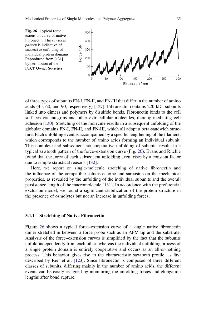
This complete and subsequent noncooperative unfolding of subunits results in a
typical sawtooth pattern of the force–extension curve (Fig. 26). Evans and Ritchie
found that the force of each subsequent unfolding event rises by a constant factor
due to simple statistical reasons [132].
Here, we report on single-molecule stretching of native fibronectin and
the influence of the compatible solutes ectoine and sarcosine on the mechanical
properties, as revealed by the unfolding of the individual subunits and the overall
persistence length of the macromolecule [131]. In accordance with the preferential
exclusion model, we found a significant stabilization of the protein structure in
the presence of osmolytes but not an increase in unfolding forces.
3.1.1 Stretching of Native Fibronectin
Figure 26 shows a typical force–extension curve of a single native fibronectin
dimer stretched in between a force probe such as an AFM tip and the substrate.
Analysis of the force–extension curves is simplified by the fact that the subunits
unfold independently from each other, whereas the individual unfolding process of
a single protein domain is entirely cooperative and occurs as an all-or-nothing
process. This behavior gives rise to the characteristic sawtooth profile, as first
described by Rief et al. [123]. Since fibronectin is composed of three different
classes of subunits, differing mainly in the number of amino acids, the different
events can be easily assigned by monitoring the unfolding forces and elongation
lengths after bond rupture.
500
400
300
200
100
0
Force / pN
300
250
200
150
100
50
0
Extension / nm
Fig. 26 Typical force
extension curve of native
fibronectin. The sawtooth
pattern is indicative of
successive unfolding of
individual protein domains.
Reproduced from [131]
by permission of the
PCCP Owner Societies
Mechanical Properties of Single Molecules and Polymer Aggregates
35
acids (45, 60, and 90, respectively) [127]. Fibronectin contains 220 kDa subunits
linked into dimers and polymers by disulfide bonds. Fibronectin binds to the cell
surfaces via integrins and other extracellular molecules, thereby mediating cell
adhesion [130]. Stretching of the molecule results in a subsequent unfolding of the
globular domains FN-I, FN-II, and FN-III, which all adopt a beta-sandwich structure. Each unfolding event is accompanied by a specific lengthening of the filament,
which corresponds to the number of amino acids forming an individual subunit.
This complete and subsequent noncooperative unfolding of subunits results in a
typical sawtooth pattern of the force–extension curve (Fig. 26). Evans and Ritchie
found that the force of each subsequent unfolding event rises by a constant factor
due to simple statistical reasons [132].
Here, we report on single-molecule stretching of native fibronectin and
the influence of the compatible solutes ectoine and sarcosine on the mechanical
properties, as revealed by the unfolding of the individual subunits and the overall
persistence length of the macromolecule [131]. In accordance with the preferential
exclusion model, we found a significant stabilization of the protein structure in
the presence of osmolytes but not an increase in unfolding forces.
3.1.1 Stretching of Native Fibronectin
Figure 26 shows a typical force–extension curve of a single native fibronectin
dimer stretched in between a force probe such as an AFM tip and the substrate.
Analysis of the force–extension curves is simplified by the fact that the subunits
unfold independently from each other, whereas the individual unfolding process of
a single protein domain is entirely cooperative and occurs as an all-or-nothing
process. This behavior gives rise to the characteristic sawtooth profile, as first
described by Rief et al. [123]. Since fibronectin is composed of three different
classes of subunits, differing mainly in the number of amino acids, the different
events can be easily assigned by monitoring the unfolding forces and elongation
lengths after bond rupture.
500
400
300
200
100
0
Force / pN
300
250
200
150
100
50
0
Extension / nm
Fig. 26 Typical force
extension curve of native
fibronectin. The sawtooth
pattern is indicative of
successive unfolding of
individual protein domains.
Reproduced from [131]
by permission of the
PCCP Owner Societies
Mechanical Properties of Single Molecules and Polymer Aggregates
35