

cyclic amino acid, is the most common solute found in the cytosol of aerobic
heterotrophic bacteria where it increases the thermotolerance [114, 115]. In general,
it has been suggested that the presence of osmolytes increases the stability of
proteins and preserves enzymatic activity in an aqueous environment. These properties make the compounds potential candidates for biotechnological applications
ranging from food processing to protection of tissue from environmental stress
factors such as UV irradiation [116]. Timasheff and coworkers [117] argue that
the main factor in stabilizing protein structure by large quantities of osmolytes
dissolved in water originates from the increase in surface tension of the water, thus
leading to a preferential exclusion of the osmolytes from the protein–water interface. The increase in surface tension is in accordance with Gibbs’ isotherm and
forces the protein to adopt a more compact protein structure that reduces the surface
area exposed to the aqueous phase without perturbing its native function.
Intrigued by this mechanism of protein stabilization, we wanted to prove this
hypothesis of protein stabilization by employing single-molecule stretching experiments using proteins. We deliberately chose proteins from the extracellular matrix
because they would be exposed to external stresses in the most profound way.
AFM and particularly single-molecule force spectroscopy can provide insights
into the stability of single ligand–receptor pairs and the elastic properties of
individual macromolecules [118–120]. Besides stretching of simple homopolymers, a variety of different biological macromolecules ranging from polysaccharides to modular proteins such as titin and spectrin have been investigated
by means of force–extension curves [118, 121, 122]. Most of the pioneering work
in this area stems from the group of Gaub and coworkers [123, 124]. Force-induced
unfolding of proteins is particular interesting for unraveling the structure–function
relationships of protein filaments involved in the mechanical function of cells and
extracellular matrix. Stretching of modular proteins such as titin [123], tenascin
[125], spectrin [126], and fibronectin [127] at different loading rates can be used to
gather information about the energy landscape of the folded structure. Thereby,
Mu ¨ller and coworkers [128] were able to remove individual domains of single
bacteriorhodopsin molecules from a solid supported membrane, giving unfolding
fingerprints displaying the structural integrity of the protein under investigation [129].
Previously, we were able to measure the mechanical unfolding of single native
fibronectin to reveal the detailed composition of the protein, known to consist
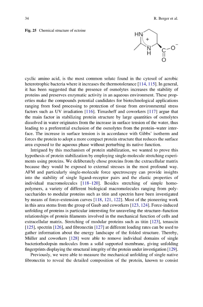
Fig. 25 Chemical structure of ectoine
34
R. Berger et al.
heterotrophic bacteria where it increases the thermotolerance [114, 115]. In general,
it has been suggested that the presence of osmolytes increases the stability of
proteins and preserves enzymatic activity in an aqueous environment. These properties make the compounds potential candidates for biotechnological applications
ranging from food processing to protection of tissue from environmental stress
factors such as UV irradiation [116]. Timasheff and coworkers [117] argue that
the main factor in stabilizing protein structure by large quantities of osmolytes
dissolved in water originates from the increase in surface tension of the water, thus
leading to a preferential exclusion of the osmolytes from the protein–water interface. The increase in surface tension is in accordance with Gibbs’ isotherm and
forces the protein to adopt a more compact protein structure that reduces the surface
area exposed to the aqueous phase without perturbing its native function.
Intrigued by this mechanism of protein stabilization, we wanted to prove this
hypothesis of protein stabilization by employing single-molecule stretching experiments using proteins. We deliberately chose proteins from the extracellular matrix
because they would be exposed to external stresses in the most profound way.
AFM and particularly single-molecule force spectroscopy can provide insights
into the stability of single ligand–receptor pairs and the elastic properties of
individual macromolecules [118–120]. Besides stretching of simple homopolymers, a variety of different biological macromolecules ranging from polysaccharides to modular proteins such as titin and spectrin have been investigated
by means of force–extension curves [118, 121, 122]. Most of the pioneering work
in this area stems from the group of Gaub and coworkers [123, 124]. Force-induced
unfolding of proteins is particular interesting for unraveling the structure–function
relationships of protein filaments involved in the mechanical function of cells and
extracellular matrix. Stretching of modular proteins such as titin [123], tenascin
[125], spectrin [126], and fibronectin [127] at different loading rates can be used to
gather information about the energy landscape of the folded structure. Thereby,
Mu ¨ller and coworkers [128] were able to remove individual domains of single
bacteriorhodopsin molecules from a solid supported membrane, giving unfolding
fingerprints displaying the structural integrity of the protein under investigation [129].
Previously, we were able to measure the mechanical unfolding of single native
fibronectin to reveal the detailed composition of the protein, known to consist
Fig. 25 Chemical structure of ectoine
34
R. Berger et al.