

toxicity and was investigated with regard to its potential to interact with subcellular
structures [20]. An interaction with the highly negatively charged extracellular matrix
(ECM) was observed at physiological pH [20]. Because the ECM surrounds cells and
plays an important role in many aspects of cellular fate (including cell migration, stem
cell differentiation, and cancer progression) specific labeling of the ECM could be
attractive for cell studies. Via affinity analysis, aggrecan was identified as a major
interaction partner [20]. Aggrecan is a major structural proteoglycan of cartilage
ECM, with a 210–250 kDa core protein to which 100–150 chondroitin sulfate and
keratan sulfate chains are attached [21, 22]. In a dot blotting assay, only negatively
charged aggrecan was found to interact with the polycationic dendrimer, most
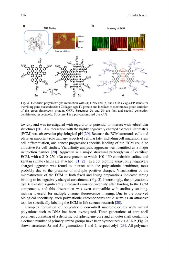
probably due to the presence of multiple positive charges. Visualization of the
microstructure of the ECM in both fixed and living preparations indicated strong
binding to its negatively charged constituents (Fig. 2). Interestingly, the polycationic
dye 4 revealed significantly increased emission intensity after binding to the ECM
components, and this observation was even compatible with antibody staining,
making it useful for multiple channel fluorescence imaging. Due to the observed
biological specificity, such polycationic chromophores could serve as an attractive
tool for specifically labeling the ECM in life science research [20].
Complex formation of polycationic core–shell macromolecules with natural
polyanions such as DNA has been investigated. Three generations of core–shell
polymers consisting of a dendritic polyphenylene core and an outer shell containing
a defined number of primary amino groups have been synthesized via ATRP (Fig. 2a
shows structures 3a and 3b, generations 1 and 2, respectively) [23]. All polymers
Fig. 2 Dendritic polyelectrolyte interaction with (a) DNA and (b) the ECM (Vkg-GFP stands for
the viking gene that codes for a Collagen type IV protein and localizes to membranes, green emission
of the green fluorescent protein, GFP). Structures 3a and 3b are first and second generation
dendrimers, respectively. Structure 4 is a polycationic red dye (P1)
216
J. Hedrich et al.
structures [20]. An interaction with the highly negatively charged extracellular matrix
(ECM) was observed at physiological pH [20]. Because the ECM surrounds cells and
plays an important role in many aspects of cellular fate (including cell migration, stem
cell differentiation, and cancer progression) specific labeling of the ECM could be
attractive for cell studies. Via affinity analysis, aggrecan was identified as a major
interaction partner [20]. Aggrecan is a major structural proteoglycan of cartilage
ECM, with a 210–250 kDa core protein to which 100–150 chondroitin sulfate and
keratan sulfate chains are attached [21, 22]. In a dot blotting assay, only negatively
charged aggrecan was found to interact with the polycationic dendrimer, most
probably due to the presence of multiple positive charges. Visualization of the
microstructure of the ECM in both fixed and living preparations indicated strong
binding to its negatively charged constituents (Fig. 2). Interestingly, the polycationic
dye 4 revealed significantly increased emission intensity after binding to the ECM
components, and this observation was even compatible with antibody staining,
making it useful for multiple channel fluorescence imaging. Due to the observed
biological specificity, such polycationic chromophores could serve as an attractive
tool for specifically labeling the ECM in life science research [20].
Complex formation of polycationic core–shell macromolecules with natural
polyanions such as DNA has been investigated. Three generations of core–shell
polymers consisting of a dendritic polyphenylene core and an outer shell containing
a defined number of primary amino groups have been synthesized via ATRP (Fig. 2a
shows structures 3a and 3b, generations 1 and 2, respectively) [23]. All polymers
Fig. 2 Dendritic polyelectrolyte interaction with (a) DNA and (b) the ECM (Vkg-GFP stands for
the viking gene that codes for a Collagen type IV protein and localizes to membranes, green emission
of the green fluorescent protein, GFP). Structures 3a and 3b are first and second generation
dendrimers, respectively. Structure 4 is a polycationic red dye (P1)
216
J. Hedrich et al.