

For further investigations, starting from a dried copolymer and Nile Red film as
described above, Nile Red-loaded polymersomes were prepared via the rehydration
method. This method leads to vesicles with very broad size distribution and
diameters ranging from 60 up to 3 μm. We imaged the fluorescently labeled
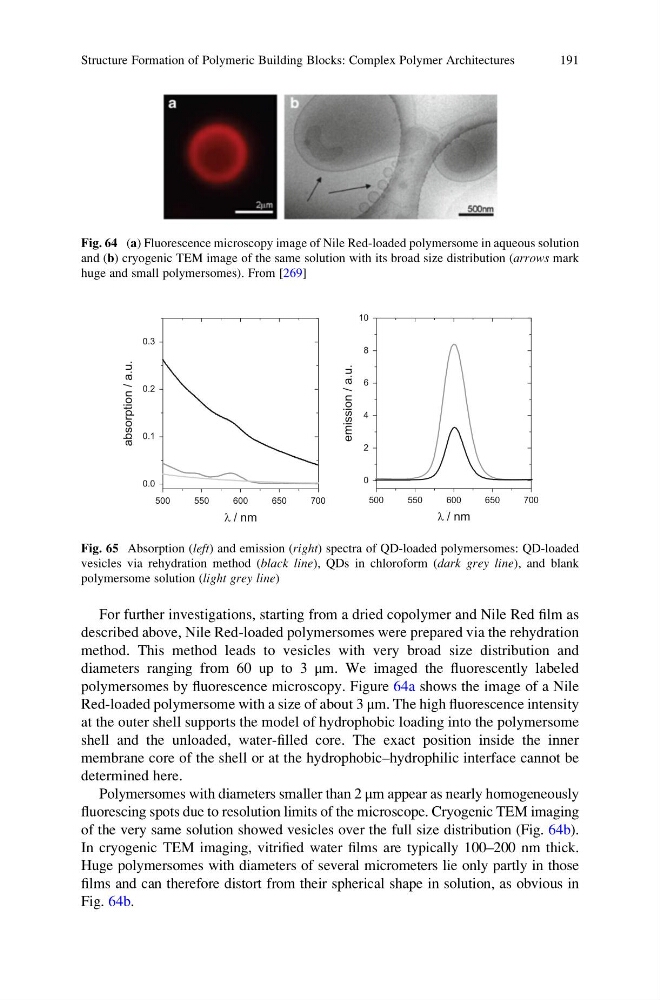
polymersomes by fluorescence microscopy. Figure 64a shows the image of a Nile
Red-loaded polymersome with a size of about 3 μm. The high fluorescence intensity
at the outer shell supports the model of hydrophobic loading into the polymersome
shell and the unloaded, water-filled core. The exact position inside the inner
membrane core of the shell or at the hydrophobic–hydrophilic interface cannot be
determined here.
Polymersomes with diameters smaller than 2 μm appear as nearly homogeneously
fluorescing spots due to resolution limits of the microscope. Cryogenic TEM imaging
of the very same solution showed vesicles over the full size distribution (Fig. 64b).
In cryogenic TEM imaging, vitrified water films are typically 100–200 nm thick.
Huge polymersomes with diameters of several micrometers lie only partly in those
films and can therefore distort from their spherical shape in solution, as obvious in
Fig. 64b.
Fig. 64 (a) Fluorescence microscopy image of Nile Red-loaded polymersome in aqueous solution
and (b) cryogenic TEM image of the same solution with its broad size distribution (arrows mark
huge and small polymersomes). From [269]
500
550
600
650
700
0.0
0.1
0.2
0.3
absorption / a.u.
l / nm
500
550
600
650
700
0
2
4
6
8
10
emission / a.u.
l / nm
Fig. 65 Absorption (left) and emission (right) spectra of QD-loaded polymersomes: QD-loaded
vesicles via rehydration method (black line), QDs in chloroform (dark grey line), and blank
polymersome solution (light grey line)
Structure Formation of Polymeric Building Blocks: Complex Polymer Architectures
191
described above, Nile Red-loaded polymersomes were prepared via the rehydration
method. This method leads to vesicles with very broad size distribution and
diameters ranging from 60 up to 3 μm. We imaged the fluorescently labeled
polymersomes by fluorescence microscopy. Figure 64a shows the image of a Nile
Red-loaded polymersome with a size of about 3 μm. The high fluorescence intensity
at the outer shell supports the model of hydrophobic loading into the polymersome
shell and the unloaded, water-filled core. The exact position inside the inner
membrane core of the shell or at the hydrophobic–hydrophilic interface cannot be
determined here.
Polymersomes with diameters smaller than 2 μm appear as nearly homogeneously
fluorescing spots due to resolution limits of the microscope. Cryogenic TEM imaging
of the very same solution showed vesicles over the full size distribution (Fig. 64b).
In cryogenic TEM imaging, vitrified water films are typically 100–200 nm thick.
Huge polymersomes with diameters of several micrometers lie only partly in those
films and can therefore distort from their spherical shape in solution, as obvious in
Fig. 64b.
Fig. 64 (a) Fluorescence microscopy image of Nile Red-loaded polymersome in aqueous solution
and (b) cryogenic TEM image of the same solution with its broad size distribution (arrows mark
huge and small polymersomes). From [269]
500
550
600
650
700
0.0
0.1
0.2
0.3
absorption / a.u.
l / nm
500
550
600
650
700
0
2
4
6
8
10
emission / a.u.
l / nm
Fig. 65 Absorption (left) and emission (right) spectra of QD-loaded polymersomes: QD-loaded
vesicles via rehydration method (black line), QDs in chloroform (dark grey line), and blank
polymersome solution (light grey line)
Structure Formation of Polymeric Building Blocks: Complex Polymer Architectures
191