

The encapsulation of a larger hydrophobic model substrate was successfully
realized utilizing highly fluorescent QDs. The enclosing of these nanoparticles inside
the PB-b-PEO polymersomes was performed only via the film rehydration procedure
because the preferred dispersant of QDs, chloroform, is immiscible with water and
therefore not suitable for the cosolvent method. The rehydration procedure for QDs
was done analogously to that for the Nile Red samples. Afterwards, filtration through
0.45-μm filters ensured a smaller vesicle size regime, which is important for further
characterization. Absorption and emission spectra indicated the presence of QDs in
the vesicle solution after filtration (Fig. 65).
The quantification of QD load in the polymersomes has not yet been possible
because the rehydration method never led to a complete solution of material and the
QD absorption peak is drowned in the high scattering background of those huge and
broadly distributed polymersomes. Furthermore, exact reference concentrations are
difficult to determine for core–shell QD solutions.
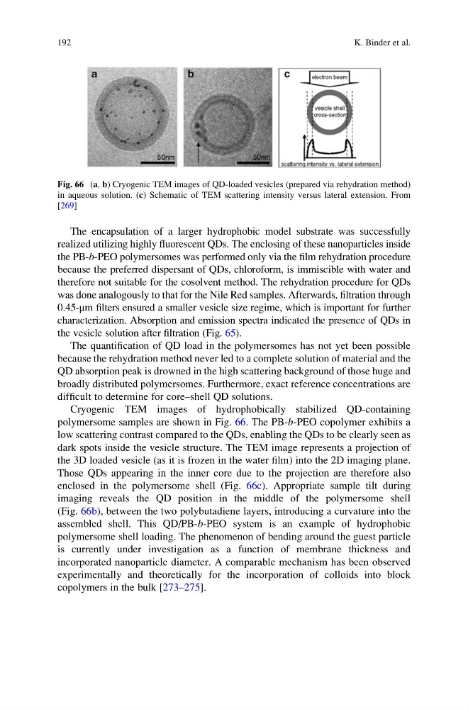
Cryogenic TEM images of hydrophobically stabilized QD-containing
polymersome samples are shown in Fig. 66. The PB-b-PEO copolymer exhibits a
low scattering contrast compared to the QDs, enabling the QDs to be clearly seen as
dark spots inside the vesicle structure. The TEM image represents a projection of
the 3D loaded vesicle (as it is frozen in the water film) into the 2D imaging plane.
Those QDs appearing in the inner core due to the projection are therefore also
enclosed in the polymersome shell (Fig. 66c). Appropriate sample tilt during
imaging reveals the QD position in the middle of the polymersome shell
(Fig. 66b), between the two polybutadiene layers, introducing a curvature into the
assembled shell. This QD/PB-b-PEO system is an example of hydrophobic
polymersome shell loading. The phenomenon of bending around the guest particle
is currently under investigation as a function of membrane thickness and
incorporated nanoparticle diameter. A comparable mechanism has been observed
experimentally and theoretically for the incorporation of colloids into block
copolymers in the bulk [273–275].
Fig. 66 (a, b) Cryogenic TEM images of QD-loaded vesicles (prepared via rehydration method)
in aqueous solution. (c) Schematic of TEM scattering intensity versus lateral extension. From
[269]
192
K. Binder et al.
realized utilizing highly fluorescent QDs. The enclosing of these nanoparticles inside
the PB-b-PEO polymersomes was performed only via the film rehydration procedure
because the preferred dispersant of QDs, chloroform, is immiscible with water and
therefore not suitable for the cosolvent method. The rehydration procedure for QDs
was done analogously to that for the Nile Red samples. Afterwards, filtration through
0.45-μm filters ensured a smaller vesicle size regime, which is important for further
characterization. Absorption and emission spectra indicated the presence of QDs in
the vesicle solution after filtration (Fig. 65).
The quantification of QD load in the polymersomes has not yet been possible
because the rehydration method never led to a complete solution of material and the
QD absorption peak is drowned in the high scattering background of those huge and
broadly distributed polymersomes. Furthermore, exact reference concentrations are
difficult to determine for core–shell QD solutions.
Cryogenic TEM images of hydrophobically stabilized QD-containing
polymersome samples are shown in Fig. 66. The PB-b-PEO copolymer exhibits a
low scattering contrast compared to the QDs, enabling the QDs to be clearly seen as
dark spots inside the vesicle structure. The TEM image represents a projection of
the 3D loaded vesicle (as it is frozen in the water film) into the 2D imaging plane.
Those QDs appearing in the inner core due to the projection are therefore also
enclosed in the polymersome shell (Fig. 66c). Appropriate sample tilt during
imaging reveals the QD position in the middle of the polymersome shell
(Fig. 66b), between the two polybutadiene layers, introducing a curvature into the
assembled shell. This QD/PB-b-PEO system is an example of hydrophobic
polymersome shell loading. The phenomenon of bending around the guest particle
is currently under investigation as a function of membrane thickness and
incorporated nanoparticle diameter. A comparable mechanism has been observed
experimentally and theoretically for the incorporation of colloids into block
copolymers in the bulk [273–275].
Fig. 66 (a, b) Cryogenic TEM images of QD-loaded vesicles (prepared via rehydration method)
in aqueous solution. (c) Schematic of TEM scattering intensity versus lateral extension. From
[269]
192
K. Binder et al.