

polymersomes was achieved by the addition of QDs to the starting copolymer/
chloroform solution at a QD/copolymer weight ratio of 1/2.8, corresponding to a
molar ratio of 1/600. To load polymersomes with Nile Red, the dye was added to
the starting copolymer/THF solution at a dye/copolymer weight ratio of 1/20.
The rehydration procedure was performed as described above.
In order to study the potential influence of the surrounding polarity on the optical
properties of an encapsulated hydrophobic dye, the hydrophobic dye Nile Red was
employed to load vesicles via the “cosolvent method” starting from a solution of
PB-b-PEO copolymer and Nile Red in THF and dropwise water addition as
described above. This leads to homogeneously colored polymersome solutions
with average hydrodynamic radii in the range of 40–50 nm. Typically, 500–550
dye molecules are taken up per vesicle, as determined from absorption spectra after
calibration with Nile Red solutions in polybutadiene. This corresponds to an uptake
of 3 mg of Nile Red per 1 g of copolymer. Blank polymersome samples without
Nile Red were prepared the same way. DLS measurements showed no differences
in size or size distribution of the polymersomes with or without Nile Red.
Cryogenic TEM images proved the vesicular structure for both cases. Analogous
to the Nile Red-loaded polymersomes, blind samples with no copolymer or with
just PEO polymer with molecular weight of approximately 3,500 g/mol were
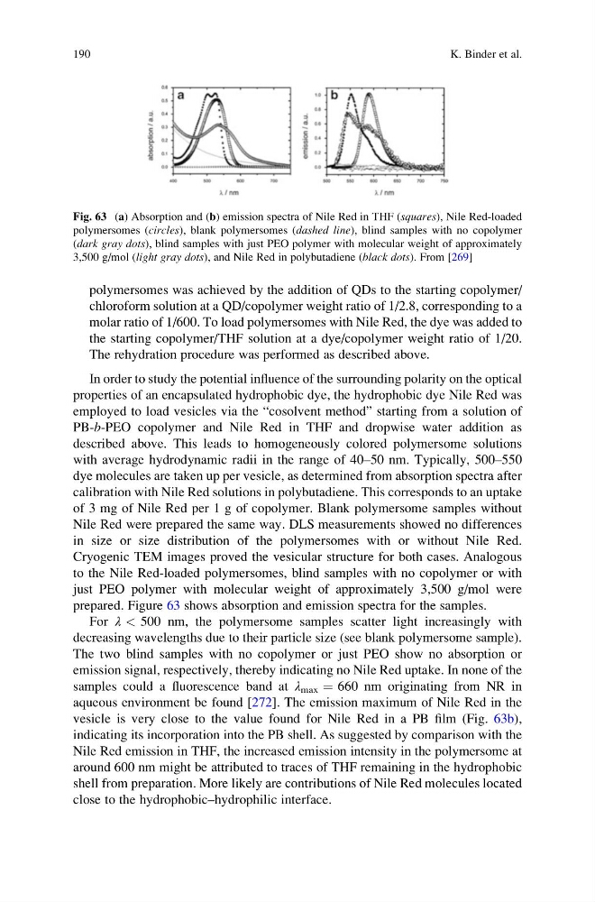
prepared. Figure 63 shows absorption and emission spectra for the samples.
For λ < 500 nm, the polymersome samples scatter light increasingly with
decreasing wavelengths due to their particle size (see blank polymersome sample).
The two blind samples with no copolymer or just PEO show no absorption or
emission signal, respectively, thereby indicating no Nile Red uptake. In none of the
samples could a fluorescence band at λ max ¼ 660 nm originating from NR in
aqueous environment be found [272]. The emission maximum of Nile Red in the
vesicle is very close to the value found for Nile Red in a PB film (Fig. 63b),
indicating its incorporation into the PB shell. As suggested by comparison with the
Nile Red emission in THF, the increased emission intensity in the polymersome at
around 600 nm might be attributed to traces of THF remaining in the hydrophobic
shell from preparation. More likely are contributions of Nile Red molecules located
close to the hydrophobic–hydrophilic interface.
Fig. 63 (a) Absorption and (b) emission spectra of Nile Red in THF (squares), Nile Red-loaded
polymersomes (circles), blank polymersomes (dashed line), blind samples with no copolymer
(dark gray dots), blind samples with just PEO polymer with molecular weight of approximately
3,500 g/mol (light gray dots), and Nile Red in polybutadiene (black dots). From [269]
190
K. Binder et al.
chloroform solution at a QD/copolymer weight ratio of 1/2.8, corresponding to a
molar ratio of 1/600. To load polymersomes with Nile Red, the dye was added to
the starting copolymer/THF solution at a dye/copolymer weight ratio of 1/20.
The rehydration procedure was performed as described above.
In order to study the potential influence of the surrounding polarity on the optical
properties of an encapsulated hydrophobic dye, the hydrophobic dye Nile Red was
employed to load vesicles via the “cosolvent method” starting from a solution of
PB-b-PEO copolymer and Nile Red in THF and dropwise water addition as
described above. This leads to homogeneously colored polymersome solutions
with average hydrodynamic radii in the range of 40–50 nm. Typically, 500–550
dye molecules are taken up per vesicle, as determined from absorption spectra after
calibration with Nile Red solutions in polybutadiene. This corresponds to an uptake
of 3 mg of Nile Red per 1 g of copolymer. Blank polymersome samples without
Nile Red were prepared the same way. DLS measurements showed no differences
in size or size distribution of the polymersomes with or without Nile Red.
Cryogenic TEM images proved the vesicular structure for both cases. Analogous
to the Nile Red-loaded polymersomes, blind samples with no copolymer or with
just PEO polymer with molecular weight of approximately 3,500 g/mol were
prepared. Figure 63 shows absorption and emission spectra for the samples.
For λ < 500 nm, the polymersome samples scatter light increasingly with
decreasing wavelengths due to their particle size (see blank polymersome sample).
The two blind samples with no copolymer or just PEO show no absorption or
emission signal, respectively, thereby indicating no Nile Red uptake. In none of the
samples could a fluorescence band at λ max ¼ 660 nm originating from NR in
aqueous environment be found [272]. The emission maximum of Nile Red in the
vesicle is very close to the value found for Nile Red in a PB film (Fig. 63b),
indicating its incorporation into the PB shell. As suggested by comparison with the
Nile Red emission in THF, the increased emission intensity in the polymersome at
around 600 nm might be attributed to traces of THF remaining in the hydrophobic
shell from preparation. More likely are contributions of Nile Red molecules located
close to the hydrophobic–hydrophilic interface.
Fig. 63 (a) Absorption and (b) emission spectra of Nile Red in THF (squares), Nile Red-loaded
polymersomes (circles), blank polymersomes (dashed line), blind samples with no copolymer
(dark gray dots), blind samples with just PEO polymer with molecular weight of approximately
3,500 g/mol (light gray dots), and Nile Red in polybutadiene (black dots). From [269]
190
K. Binder et al.