

passes through the “unstable regime” in which phase separation is known to occur.
Rapid mixing and vigorous stirring might avoid macroscopic phase separation, but
the problem is obvious.
Recently, a light scattering stopped-flow cell was developed with a dead time of
less than 5 ms, thus allowing for kinetic measurements of the apparent molar mass
starting at 10–50 ms after mixing [151]. With such a device, the complexation
kinetics of DNA with a cylindrical brush polymer with PVP side chains was
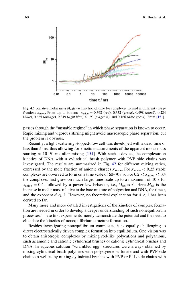
investigated. The results are summarized in Fig. 42 for different mixing ratios,
expressed by the mole fraction of anionic charges x anion . For x anion < 0.25 stable
complexes are observed to form on a time scale of 60–70 ms. For 0.2 < x anion < 0.4
the complexes first grow on much larger time scale up to a maximum of 10 s for
x anion ¼ 0.4, followed by a power law behavior, i.e., M rel % t
d
. Here M rel is the
increase in molar mass relative to the bare mixture of polycation and DNA, the time t,
and the exponent d ( 1. However, no theoretical explanation for d < 1 has been
derived so far.
Many more and more detailed investigations of the kinetics of complex formation are needed in order to develop a deeper understanding of such nonequilibrium
processes. These first experiments merely demonstrate the potential and the need to
elucidate the kinetics of nonequilibrium structure formation.
Besides investigating nonequilibrium complexes, it is equally challenging to
direct electrostatically driven complex formation into equilibrium. One vision was
to obtain anisotropic complexes by mixing rod-like polycations and polyanions,
such as anionic and cationic cylindrical brushes or cationic cylindrical brushes and
DNA. In aqueous solution “scrambled egg” structures were always obtained by
mixing cylindrical brush polymers with polystyrene sulfonate and with PVP side
chains as well as by mixing cylindrical brushes with PVP or PLL side chains with
Fig. 42 Relative molar mass M rel (t) as function of time for complexes formed at different charge
fractions x anion . From top to bottom: x anion ¼ 0.398 (red), 0.332 (green), 0.498 (black), 0.284
(blue), 0.665 (orange), 0.249 (light blue), 0.199 (magenta), and 0.166 (dark green). From [151]
160
K. Binder et al.
Rapid mixing and vigorous stirring might avoid macroscopic phase separation, but
the problem is obvious.
Recently, a light scattering stopped-flow cell was developed with a dead time of
less than 5 ms, thus allowing for kinetic measurements of the apparent molar mass
starting at 10–50 ms after mixing [151]. With such a device, the complexation
kinetics of DNA with a cylindrical brush polymer with PVP side chains was
investigated. The results are summarized in Fig. 42 for different mixing ratios,
expressed by the mole fraction of anionic charges x anion . For x anion < 0.25 stable
complexes are observed to form on a time scale of 60–70 ms. For 0.2 < x anion < 0.4
the complexes first grow on much larger time scale up to a maximum of 10 s for
x anion ¼ 0.4, followed by a power law behavior, i.e., M rel % t
d
. Here M rel is the
increase in molar mass relative to the bare mixture of polycation and DNA, the time t,
and the exponent d ( 1. However, no theoretical explanation for d < 1 has been
derived so far.
Many more and more detailed investigations of the kinetics of complex formation are needed in order to develop a deeper understanding of such nonequilibrium
processes. These first experiments merely demonstrate the potential and the need to
elucidate the kinetics of nonequilibrium structure formation.
Besides investigating nonequilibrium complexes, it is equally challenging to
direct electrostatically driven complex formation into equilibrium. One vision was
to obtain anisotropic complexes by mixing rod-like polycations and polyanions,
such as anionic and cationic cylindrical brushes or cationic cylindrical brushes and
DNA. In aqueous solution “scrambled egg” structures were always obtained by
mixing cylindrical brush polymers with polystyrene sulfonate and with PVP side
chains as well as by mixing cylindrical brushes with PVP or PLL side chains with
Fig. 42 Relative molar mass M rel (t) as function of time for complexes formed at different charge
fractions x anion . From top to bottom: x anion ¼ 0.398 (red), 0.332 (green), 0.498 (black), 0.284
(blue), 0.665 (orange), 0.249 (light blue), 0.199 (magenta), and 0.166 (dark green). From [151]
160
K. Binder et al.