

materials (BSM) or tissue engineering scaffolds (TES). Recently, we reported on
PEC nanoparticles that were loaded by the bisphosphonate pamidronate (PAM) and
deposited as adhesive films onto planar Ge model substrates [149].
Bisphosphonates like PAM are known to inhibit osteoclastic activity via the
farnesyl pathway [150], favoring osteoblastic bone formation, and are widely
used as therapeutics for systemic bone diseases like osteoporosis [151]. A retarded
release of PAM under conservation and adhesive stability of the bare PEC particle
film was shown by in situ ATR-FTIR spectroscopy, monitoring the depletion of
PAM in the cast PEC film matrix. Up to now, this surface-sensitive method has
been widely applied by us to analyze the sorption and conformation processes of
PEC layers [152, 153], melanin-like films [154], chiral model drug compounds
[155], lipids [156], peptides [157], and proteins [158]. Various factors of PAM
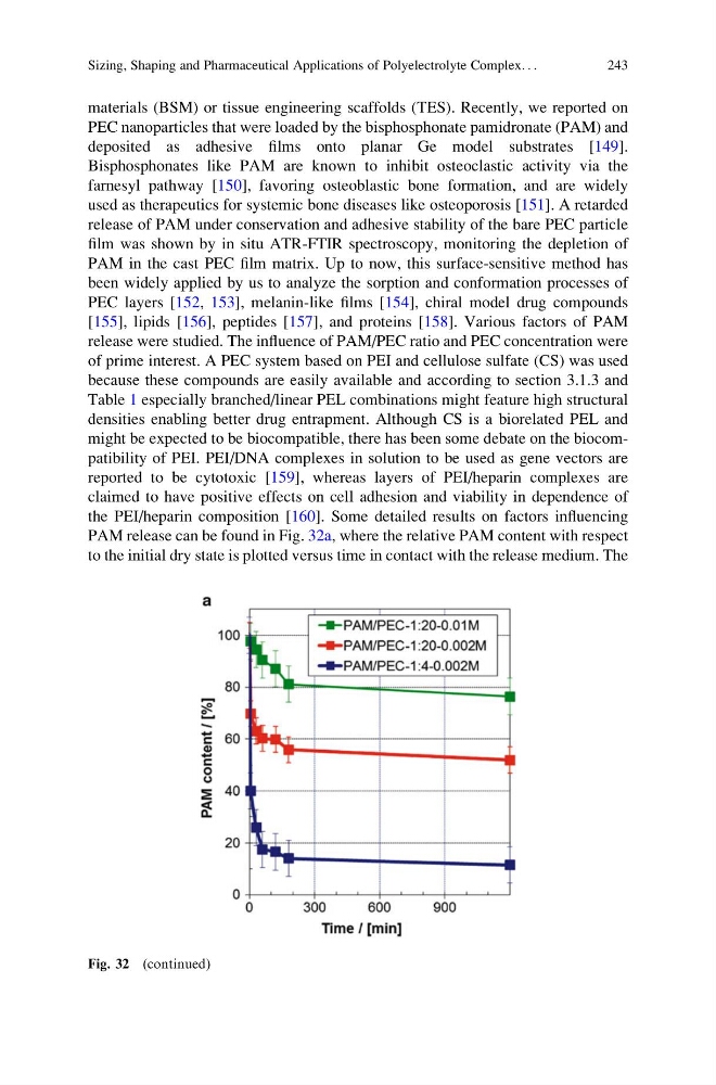
release were studied. The influence of PAM/PEC ratio and PEC concentration were
of prime interest. A PEC system based on PEI and cellulose sulfate (CS) was used
because these compounds are easily available and according to section 3.1.3 and
Table 1 especially branched/linear PEL combinations might feature high structural
densities enabling better drug entrapment. Although CS is a biorelated PEL and
might be expected to be biocompatible, there has been some debate on the biocompatibility of PEI. PEI/DNA complexes in solution to be used as gene vectors are
reported to be cytotoxic [159], whereas layers of PEI/heparin complexes are
claimed to have positive effects on cell adhesion and viability in dependence of
the PEI/heparin composition [160]. Some detailed results on factors influencing
PAM release can be found in Fig. 32a, where the relative PAM content with respect
to the initial dry state is plotted versus time in contact with the release medium. The
Fig. 32 (continued)
Sizing, Shaping and Pharmaceutical Applications of Polyelectrolyte Complex. . .
243
PEC nanoparticles that were loaded by the bisphosphonate pamidronate (PAM) and
deposited as adhesive films onto planar Ge model substrates [149].
Bisphosphonates like PAM are known to inhibit osteoclastic activity via the
farnesyl pathway [150], favoring osteoblastic bone formation, and are widely
used as therapeutics for systemic bone diseases like osteoporosis [151]. A retarded
release of PAM under conservation and adhesive stability of the bare PEC particle
film was shown by in situ ATR-FTIR spectroscopy, monitoring the depletion of
PAM in the cast PEC film matrix. Up to now, this surface-sensitive method has
been widely applied by us to analyze the sorption and conformation processes of
PEC layers [152, 153], melanin-like films [154], chiral model drug compounds
[155], lipids [156], peptides [157], and proteins [158]. Various factors of PAM
release were studied. The influence of PAM/PEC ratio and PEC concentration were
of prime interest. A PEC system based on PEI and cellulose sulfate (CS) was used
because these compounds are easily available and according to section 3.1.3 and
Table 1 especially branched/linear PEL combinations might feature high structural
densities enabling better drug entrapment. Although CS is a biorelated PEL and
might be expected to be biocompatible, there has been some debate on the biocompatibility of PEI. PEI/DNA complexes in solution to be used as gene vectors are
reported to be cytotoxic [159], whereas layers of PEI/heparin complexes are
claimed to have positive effects on cell adhesion and viability in dependence of
the PEI/heparin composition [160]. Some detailed results on factors influencing
PAM release can be found in Fig. 32a, where the relative PAM content with respect
to the initial dry state is plotted versus time in contact with the release medium. The
Fig. 32 (continued)
Sizing, Shaping and Pharmaceutical Applications of Polyelectrolyte Complex. . .
243