

In cardiovascular devices, it is essential to impart hemocompatibility to PVA-C.
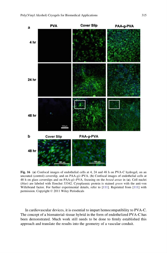
The concept of a biomaterial–tissue hybrid in the form of endothelized PVA-C has
been demonstrated. Much work still needs to be done to firmly established this
approach and translate the results into the geometry of a vascular conduit.
Fig. 16 (a) Confocal images of endothelial cells at 4, 24 and 48 h on PVA-C hydrogel, on an
uncoated (control) coverslip, and on PAA-g/c-PVA. (b) Confocal images of endothelial cells at
48 h on glass coverslips and on PAA-g/c-PVA, focusing on the boxed areas in (a). Cell nuclei
(blue) are labeled with Hoechst 33342. Cytoplasmic protein is stained green with the anti-von
Willebrand factor. For further experimental details, refer to [111]. Reprinted from [111] with
permission. Copyright © 2011 Wiley Periodicals
Poly(Vinyl Alcohol) Cryogels for Biomedical Applications
315
The concept of a biomaterial–tissue hybrid in the form of endothelized PVA-C has
been demonstrated. Much work still needs to be done to firmly established this
approach and translate the results into the geometry of a vascular conduit.
Fig. 16 (a) Confocal images of endothelial cells at 4, 24 and 48 h on PVA-C hydrogel, on an
uncoated (control) coverslip, and on PAA-g/c-PVA. (b) Confocal images of endothelial cells at
48 h on glass coverslips and on PAA-g/c-PVA, focusing on the boxed areas in (a). Cell nuclei
(blue) are labeled with Hoechst 33342. Cytoplasmic protein is stained green with the anti-von
Willebrand factor. For further experimental details, refer to [111]. Reprinted from [111] with
permission. Copyright © 2011 Wiley Periodicals
Poly(Vinyl Alcohol) Cryogels for Biomedical Applications
315