

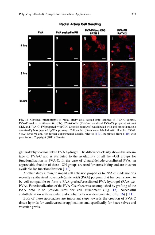
glutaraldehyde-crosslinked PVA hydrogel. The difference clearly shows the advantage of PVA-C and is attributed to the availability of all the –OH groups for
functionalization in PVA-C. In the case of glutaraldehyde-crosslinked PVA, an
appreciable fraction of these –OH groups are used for crosslinking and are thus not
available for functionalization [110].
Another study aiming to impart cell adhesion properties to PVA-C made use of a
recently synthesized novel poly(amic acid) (PAA) polymer that has been shown to
be cell compatible to form a PAA-grafted/crosslinked-PVA hydrogel (PAA-g/cPVA). Functionalization of the PVA-C surface was accomplished by grafting of the
PAA onto it to provide sites for cell attachment (Fig. 15). Successful
endothelization with vascular endothelial cells was demonstrated (Fig. 16) [111].
Both of these approaches are important steps towards the creation of PVA-C
tissue hybrids for cardiovascular applications and specifically for heart valves and
vascular grafts.
Fig. 14 Confocal micrographs of radial artery cells seeded onto samples of PVA-C control,
PVA-C soaked in fibronectin (FN), PVA-C–FN (FN-functionalized PVA-C) prepared without
CDI, and PVA-C–FN prepared with CDI. Cytoskeleton (red) was labeled with anti-smooth muscle
α-actin–Cy3-conjugated IgG2a primary. Cell nuclei (blue) were labeled with Hoechst 33342.
Scale bars: 50 μm. For further experimental details, refer to [110]. Reprinted from [110] with
permission. Copyright (2011) Elsevier
Poly(Vinyl Alcohol) Cryogels for Biomedical Applications
313
functionalization in PVA-C. In the case of glutaraldehyde-crosslinked PVA, an
appreciable fraction of these –OH groups are used for crosslinking and are thus not
available for functionalization [110].
Another study aiming to impart cell adhesion properties to PVA-C made use of a
recently synthesized novel poly(amic acid) (PAA) polymer that has been shown to
be cell compatible to form a PAA-grafted/crosslinked-PVA hydrogel (PAA-g/cPVA). Functionalization of the PVA-C surface was accomplished by grafting of the
PAA onto it to provide sites for cell attachment (Fig. 15). Successful
endothelization with vascular endothelial cells was demonstrated (Fig. 16) [111].
Both of these approaches are important steps towards the creation of PVA-C
tissue hybrids for cardiovascular applications and specifically for heart valves and
vascular grafts.
Fig. 14 Confocal micrographs of radial artery cells seeded onto samples of PVA-C control,
PVA-C soaked in fibronectin (FN), PVA-C–FN (FN-functionalized PVA-C) prepared without
CDI, and PVA-C–FN prepared with CDI. Cytoskeleton (red) was labeled with anti-smooth muscle
α-actin–Cy3-conjugated IgG2a primary. Cell nuclei (blue) were labeled with Hoechst 33342.
Scale bars: 50 μm. For further experimental details, refer to [110]. Reprinted from [110] with
permission. Copyright (2011) Elsevier
Poly(Vinyl Alcohol) Cryogels for Biomedical Applications
313