

have direct blood contact, PVA-C would also have to be hemocompatible. Unfortunately, as with most synthetic materials, PVA is not hemocompatible [102]. One
way to overcome this is to create a hemocompatible surface for PVA-C. In vascular
tissue, hemocompatability is provided by a monolayer of vascular endothelial cell
on the tissue surface. PVA, being a very hydrophilic polymer, is not conducive to
cell adhesion [103]. Many approaches have been tried to promote cell adhesion to
the PVA surface with varying degrees of success [79, 104–109]. We have recently
reported two approaches that successfully functionalized the PVA-C surface for
cell adhesion, including the vascular endothelial cells that are required for
hemocompatibility. The resulting material consists of mechanically tuned PVA-C
and a living interface of vascular endothelial cells. It can be regarded as belonging
to a novel class of “biomaterial–tissue” hybrid materials and, in the present
combination of materials, we called it a “PVA-C tissue hybrid”.
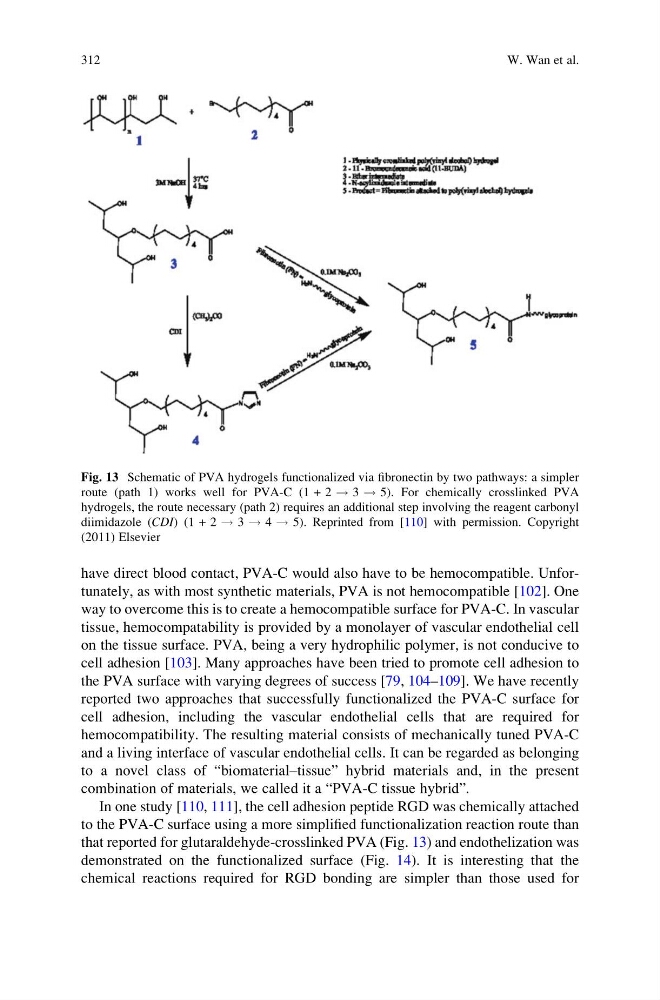
In one study [110, 111], the cell adhesion peptide RGD was chemically attached
to the PVA-C surface using a more simplified functionalization reaction route than
that reported for glutaraldehyde-crosslinked PVA (Fig. 13) and endothelization was
demonstrated on the functionalized surface (Fig. 14). It is interesting that the
chemical reactions required for RGD bonding are simpler than those used for
Fig. 13 Schematic of PVA hydrogels functionalized via fibronectin by two pathways: a simpler
route (path 1) works well for PVA-C (1 + 2 ! 3 ! 5). For chemically crosslinked PVA
hydrogels, the route necessary (path 2) requires an additional step involving the reagent carbonyl
diimidazole (CDI) (1 + 2 ! 3 ! 4 ! 5). Reprinted from [110] with permission. Copyright
(2011) Elsevier
312
W. Wan et al.
way to overcome this is to create a hemocompatible surface for PVA-C. In vascular
tissue, hemocompatability is provided by a monolayer of vascular endothelial cell
on the tissue surface. PVA, being a very hydrophilic polymer, is not conducive to
cell adhesion [103]. Many approaches have been tried to promote cell adhesion to
the PVA surface with varying degrees of success [79, 104–109]. We have recently
reported two approaches that successfully functionalized the PVA-C surface for
cell adhesion, including the vascular endothelial cells that are required for
hemocompatibility. The resulting material consists of mechanically tuned PVA-C
and a living interface of vascular endothelial cells. It can be regarded as belonging
to a novel class of “biomaterial–tissue” hybrid materials and, in the present
combination of materials, we called it a “PVA-C tissue hybrid”.
In one study [110, 111], the cell adhesion peptide RGD was chemically attached
to the PVA-C surface using a more simplified functionalization reaction route than
that reported for glutaraldehyde-crosslinked PVA (Fig. 13) and endothelization was
demonstrated on the functionalized surface (Fig. 14). It is interesting that the
chemical reactions required for RGD bonding are simpler than those used for
Fig. 13 Schematic of PVA hydrogels functionalized via fibronectin by two pathways: a simpler
route (path 1) works well for PVA-C (1 + 2 ! 3 ! 5). For chemically crosslinked PVA
hydrogels, the route necessary (path 2) requires an additional step involving the reagent carbonyl
diimidazole (CDI) (1 + 2 ! 3 ! 4 ! 5). Reprinted from [110] with permission. Copyright
(2011) Elsevier
312
W. Wan et al.