

the bioprosthetic heart valve [7]. They subjected PVA solution with 15 wt% to 1–6
FTCs with a hold time of 6 h at À20
C and thawing rate of 0.2
C/min. The
samples were tested under a constant strain rate of 40 mm/s (~200 %/s) to a
maximum of 80 % strain at 37
C. In addition, the samples were also subjected to
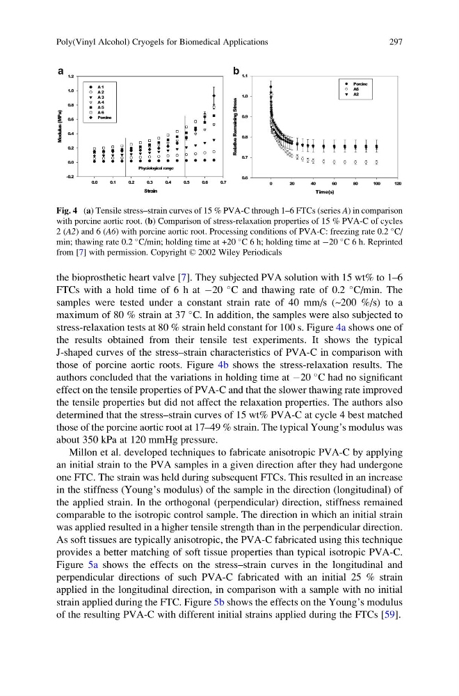
stress-relaxation tests at 80 % strain held constant for 100 s. Figure 4a shows one of
the results obtained from their tensile test experiments. It shows the typical
J-shaped curves of the stress–strain characteristics of PVA-C in comparison with
those of porcine aortic roots. Figure 4b shows the stress-relaxation results. The
authors concluded that the variations in holding time at À20
C had no significant
effect on the tensile properties of PVA-C and that the slower thawing rate improved
the tensile properties but did not affect the relaxation properties. The authors also
determined that the stress–strain curves of 15 wt% PVA-C at cycle 4 best matched
those of the porcine aortic root at 17–49 % strain. The typical Young’s modulus was
about 350 kPa at 120 mmHg pressure.
Millon et al. developed techniques to fabricate anisotropic PVA-C by applying
an initial strain to the PVA samples in a given direction after they had undergone
one FTC. The strain was held during subsequent FTCs. This resulted in an increase
in the stiffness (Young’s modulus) of the sample in the direction (longitudinal) of
the applied strain. In the orthogonal (perpendicular) direction, stiffness remained
comparable to the isotropic control sample. The direction in which an initial strain
was applied resulted in a higher tensile strength than in the perpendicular direction.
As soft tissues are typically anisotropic, the PVA-C fabricated using this technique
provides a better matching of soft tissue properties than typical isotropic PVA-C.
Figure 5a shows the effects on the stress–strain curves in the longitudinal and
perpendicular directions of such PVA-C fabricated with an initial 25 % strain
applied in the longitudinal direction, in comparison with a sample with no initial
strain applied during the FTC. Figure 5b shows the effects on the Young’s modulus
of the resulting PVA-C with different initial strains applied during the FTCs [59].
b
a
Fig. 4 (a) Tensile stress–strain curves of 15 % PVA-C through 1–6 FTCs (series A) in comparison
with porcine aortic root. (b) Comparison of stress-relaxation properties of 15 % PVA-C of cycles
2 (A2) and 6 (A6) with porcine aortic root. Processing conditions of PVA-C: freezing rate 0.2
C/
min; thawing rate 0.2
C/min; holding time at +20
C 6 h; holding time at À20
C 6 h. Reprinted
from [7] with permission. Copyright © 2002 Wiley Periodicals
Poly(Vinyl Alcohol) Cryogels for Biomedical Applications
297
FTCs with a hold time of 6 h at À20
C and thawing rate of 0.2
C/min. The
samples were tested under a constant strain rate of 40 mm/s (~200 %/s) to a
maximum of 80 % strain at 37
C. In addition, the samples were also subjected to
stress-relaxation tests at 80 % strain held constant for 100 s. Figure 4a shows one of
the results obtained from their tensile test experiments. It shows the typical
J-shaped curves of the stress–strain characteristics of PVA-C in comparison with
those of porcine aortic roots. Figure 4b shows the stress-relaxation results. The
authors concluded that the variations in holding time at À20
C had no significant
effect on the tensile properties of PVA-C and that the slower thawing rate improved
the tensile properties but did not affect the relaxation properties. The authors also
determined that the stress–strain curves of 15 wt% PVA-C at cycle 4 best matched
those of the porcine aortic root at 17–49 % strain. The typical Young’s modulus was
about 350 kPa at 120 mmHg pressure.
Millon et al. developed techniques to fabricate anisotropic PVA-C by applying
an initial strain to the PVA samples in a given direction after they had undergone
one FTC. The strain was held during subsequent FTCs. This resulted in an increase
in the stiffness (Young’s modulus) of the sample in the direction (longitudinal) of
the applied strain. In the orthogonal (perpendicular) direction, stiffness remained
comparable to the isotropic control sample. The direction in which an initial strain
was applied resulted in a higher tensile strength than in the perpendicular direction.
As soft tissues are typically anisotropic, the PVA-C fabricated using this technique
provides a better matching of soft tissue properties than typical isotropic PVA-C.
Figure 5a shows the effects on the stress–strain curves in the longitudinal and
perpendicular directions of such PVA-C fabricated with an initial 25 % strain
applied in the longitudinal direction, in comparison with a sample with no initial
strain applied during the FTC. Figure 5b shows the effects on the Young’s modulus
of the resulting PVA-C with different initial strains applied during the FTCs [59].
b
a
Fig. 4 (a) Tensile stress–strain curves of 15 % PVA-C through 1–6 FTCs (series A) in comparison
with porcine aortic root. (b) Comparison of stress-relaxation properties of 15 % PVA-C of cycles
2 (A2) and 6 (A6) with porcine aortic root. Processing conditions of PVA-C: freezing rate 0.2
C/
min; thawing rate 0.2
C/min; holding time at +20
C 6 h; holding time at À20
C 6 h. Reprinted
from [7] with permission. Copyright © 2002 Wiley Periodicals
Poly(Vinyl Alcohol) Cryogels for Biomedical Applications
297