

3.2 Nanoparticles Prepared by Reprecipitation
3.2.1 MEH-PPV Nanoparticles
The nanoscale and solid-state optical properties of MEH-PPV NPs fabricated by
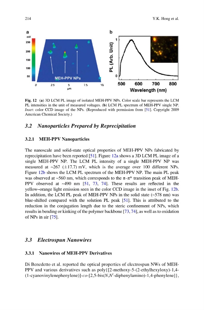
reprecipitation have been reported [51]. Figure 12a shows a 3D LCM PL image of a
single MEH-PPV NP. The LCM PL intensity of a single MEH-PPV NP was
measured at ~267 (Æ17.7) mV, which is the average over 100 different NPs.
Figure 12b shows the LCM PL spectrum of the MEH-PPV NP. The main PL peak
was observed at ~560 nm, which corresponds to the π–π* transition peak of MEHPPV observed at ~490 nm [51, 73, 74]. These results are reflected in the
yellow–orange light emission seen in the color CCD image in the inset of Fig. 12b.
In addition, the LCM PL peak of MEH-PPV NPs in the solid state (~578 nm) was
blue-shifted compared with the solution PL peak [51]. This is attributed to the
reduction in the conjugation length due to the steric confinement of NPs, which
results in bending or kinking of the polymer backbone [73, 74], as well as to oxidation
of NPs in air [75].
3.3 Electrospun Nanowires
3.3.1 Nanowires of MEH-PPV Derivatives
Di Benedetto et al. reported the optical properties of electrospun NWs of MEHPPV and various derivatives such as poly{[2-methoxy-5-(2-ethylhexyloxy)-1,4(1-cyanovinylenephenylene)]-co-[2,5-bis(N,N
0 -diphenylamino)-1,4-phenylene]},
Fig. 12 (a) 3D LCM PL image of isolated MEH-PPV NPs. Color scale bar represents the LCM
PL intensities in the unit of measured voltages. (b) LCM PL spectrum of MEH-PPV single NP.
Inset: color CCD image of the NPs. (Reproduced with permission from [51]. Copyright 2009
American Chemical Society.)
214
Y.K. Hong et al.
3.2.1 MEH-PPV Nanoparticles
The nanoscale and solid-state optical properties of MEH-PPV NPs fabricated by
reprecipitation have been reported [51]. Figure 12a shows a 3D LCM PL image of a
single MEH-PPV NP. The LCM PL intensity of a single MEH-PPV NP was
measured at ~267 (Æ17.7) mV, which is the average over 100 different NPs.
Figure 12b shows the LCM PL spectrum of the MEH-PPV NP. The main PL peak
was observed at ~560 nm, which corresponds to the π–π* transition peak of MEHPPV observed at ~490 nm [51, 73, 74]. These results are reflected in the
yellow–orange light emission seen in the color CCD image in the inset of Fig. 12b.
In addition, the LCM PL peak of MEH-PPV NPs in the solid state (~578 nm) was
blue-shifted compared with the solution PL peak [51]. This is attributed to the
reduction in the conjugation length due to the steric confinement of NPs, which
results in bending or kinking of the polymer backbone [73, 74], as well as to oxidation
of NPs in air [75].
3.3 Electrospun Nanowires
3.3.1 Nanowires of MEH-PPV Derivatives
Di Benedetto et al. reported the optical properties of electrospun NWs of MEHPPV and various derivatives such as poly{[2-methoxy-5-(2-ethylhexyloxy)-1,4(1-cyanovinylenephenylene)]-co-[2,5-bis(N,N
0 -diphenylamino)-1,4-phenylene]},
Fig. 12 (a) 3D LCM PL image of isolated MEH-PPV NPs. Color scale bar represents the LCM
PL intensities in the unit of measured voltages. (b) LCM PL spectrum of MEH-PPV single NP.
Inset: color CCD image of the NPs. (Reproduced with permission from [51]. Copyright 2009
American Chemical Society.)
214
Y.K. Hong et al.