

3 Optical Properties of As-Prepared Nanostructures
In this section, we discuss the optical properties of the as-prepared light-emitting
polymer nanostructures described in Sect. 2. The ultraviolet and visible (UV–vis)
absorption and photoluminescence (PL) spectra of the polymer nanostructures dispersed in organic solvents are introduced and discussed. The nanoscale and solid-state
optical properties of a single unit of the light-emitting polymer nanostructures can be
investigated using high-resolution laser confocal microscope (LCM) systems built
around an inverted optical microscope, coupled with luminescence color chargecoupled device (CCD) measurements. In this method, the LCM PL intensities of
single units of nanostructures are measured in units of voltage or photon count. For a
quantitative comparison of the nanoscale and solid-state optical properties of polymer
nanostructures, the LCM PL spectra must be normalized with respect to those of
pristine (i.e., as-prepared) samples. The details of the methods for the LCM and CCD
experiments are reported elsewhere [41, 42, 61–65].
3.1 Electrochemically Synthesized Nanotubes and Nanowires
3.1.1 P3MT Nanotubes
Park and coworkers reported that the doping level and structural properties of
electrochemically synthesized P3MT NTs could be controlled by varying the
synthetic temperature [40]. Figure 8a shows a comparison of the UV–vis absorption
spectra of P3MT NTs synthesized at various temperatures. For HF-treated P3MT
NTs, the π–π* transition peak shifted from 2.27 to 2.33 eV for NTs synthesized at
20
C and À20
C, respectively. Furthermore, as the synthetic temperature decreased
from 20
C to À20
C, the intensity of the bipolaron peaks at ~1.6 eV increased,
implying a variation in the degree of doping with temperature [24]. For NaOHtreated P3MT NTs, a bipolaron peak was only observed at ~1.6 eV for the NTs
synthesized at À20
C, as shown in the inset of Fig. 8a. The π–π* transition peak of
the NaOH-treated P3MT NTs was observed at ~2.15 eV. The results indicate that
the optical properties of P3MT NTs can be controlled by varying the synthetic
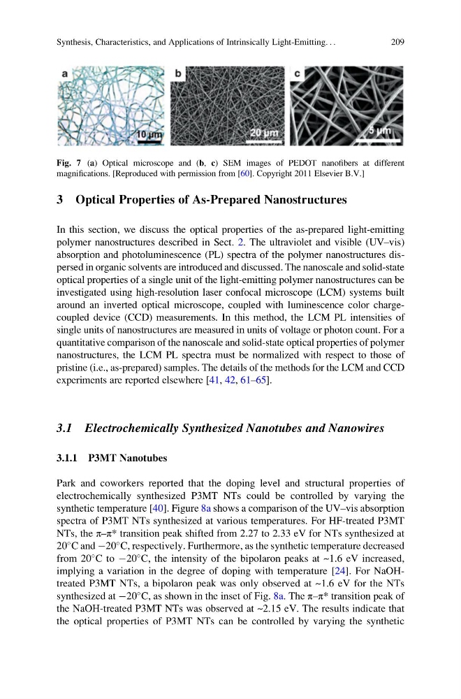
Fig. 7 (a) Optical microscope and (b, c) SEM images of PEDOT nanofibers at different
magnifications. [Reproduced with permission from [60]. Copyright 2011 Elsevier B.V.]
Synthesis, Characteristics, and Applications of Intrinsically Light-Emitting. . .
209
In this section, we discuss the optical properties of the as-prepared light-emitting
polymer nanostructures described in Sect. 2. The ultraviolet and visible (UV–vis)
absorption and photoluminescence (PL) spectra of the polymer nanostructures dispersed in organic solvents are introduced and discussed. The nanoscale and solid-state
optical properties of a single unit of the light-emitting polymer nanostructures can be
investigated using high-resolution laser confocal microscope (LCM) systems built
around an inverted optical microscope, coupled with luminescence color chargecoupled device (CCD) measurements. In this method, the LCM PL intensities of
single units of nanostructures are measured in units of voltage or photon count. For a
quantitative comparison of the nanoscale and solid-state optical properties of polymer
nanostructures, the LCM PL spectra must be normalized with respect to those of
pristine (i.e., as-prepared) samples. The details of the methods for the LCM and CCD
experiments are reported elsewhere [41, 42, 61–65].
3.1 Electrochemically Synthesized Nanotubes and Nanowires
3.1.1 P3MT Nanotubes
Park and coworkers reported that the doping level and structural properties of
electrochemically synthesized P3MT NTs could be controlled by varying the
synthetic temperature [40]. Figure 8a shows a comparison of the UV–vis absorption
spectra of P3MT NTs synthesized at various temperatures. For HF-treated P3MT
NTs, the π–π* transition peak shifted from 2.27 to 2.33 eV for NTs synthesized at
20
C and À20
C, respectively. Furthermore, as the synthetic temperature decreased
from 20
C to À20
C, the intensity of the bipolaron peaks at ~1.6 eV increased,
implying a variation in the degree of doping with temperature [24]. For NaOHtreated P3MT NTs, a bipolaron peak was only observed at ~1.6 eV for the NTs
synthesized at À20
C, as shown in the inset of Fig. 8a. The π–π* transition peak of
the NaOH-treated P3MT NTs was observed at ~2.15 eV. The results indicate that
the optical properties of P3MT NTs can be controlled by varying the synthetic
Fig. 7 (a) Optical microscope and (b, c) SEM images of PEDOT nanofibers at different
magnifications. [Reproduced with permission from [60]. Copyright 2011 Elsevier B.V.]
Synthesis, Characteristics, and Applications of Intrinsically Light-Emitting. . .
209