

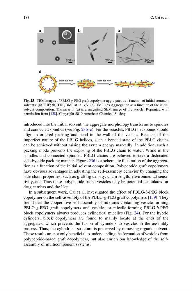
introduced into the initial solvent, the aggregate morphology transforms to spindles
and connected spindles (see Fig. 23b–c). For the vesicles, PBLG backbones should
align in ordered packing and bend in the wall of the vesicle. Because of the
imperfect nature of the PBLG helices, such a bended state of the PBLG chains
can be achieved without raising the system energy markedly. In addition, such a
packing mode prevents the exposing of the PBLG chain to water. While in the
spindles and connected spindles, PBLG chains are believed to take a dislocated
side-by-side packing manner. Figure 23d is a schematic illustration of the aggregation as a function of the initial solvent composition. Polypeptide graft copolymers
have obvious advantages in adjusting the self-assembly behavior by changing the
side-chain properties, such as grafting density, chain length, environmental sensitivity, etc. Thus these polypeptide-based vesicles may be potential candidates for
drug carriers and the like.
In a subsequent work, Cai et al. investigated the effect of PBLG-b-PEG block
copolymer on the self-assembly of the PBLG-g-PEG graft copolymers [139]. They
found that the cooperative self-assembly of mixtures containing vesicle-forming
PBLG-g-PEG graft copolymers and vesicle- or micelle-forming PBLG-b-PEG
block copolymers always produces cylindrical micelles (Fig. 24). For the hybrid
cylinders, block copolymers are found to mainly locate at the ends of the
aggregates, which prevents the fusion of cylinders to vesicles in the assembly
process. Thus, the cylindrical structure is preserved by removing organic solvent.
These results are not only beneficial to understanding the formation of vesicles from
polypeptide-based graft copolymers, but also enrich our knowledge of the selfassembly of multicomponent systems.
Increase fDMF
Increase fDMF
a
d
b
c
Fig. 23 TEM images of PBLG-g-PEG graft copolymer aggregates as a function of initial common
solvents: (a) THF; (b) THF/DMF at 1/1 v/v; (c) DMF. (d) Aggregation as a function of the initial
solvent composition. The inset in (a) is a magnified SEM image of the vesicle. Reprinted with
permission from [138]. Copyright 2010 American Chemical Society
188
C. Cai et al.
and connected spindles (see Fig. 23b–c). For the vesicles, PBLG backbones should
align in ordered packing and bend in the wall of the vesicle. Because of the
imperfect nature of the PBLG helices, such a bended state of the PBLG chains
can be achieved without raising the system energy markedly. In addition, such a
packing mode prevents the exposing of the PBLG chain to water. While in the
spindles and connected spindles, PBLG chains are believed to take a dislocated
side-by-side packing manner. Figure 23d is a schematic illustration of the aggregation as a function of the initial solvent composition. Polypeptide graft copolymers
have obvious advantages in adjusting the self-assembly behavior by changing the
side-chain properties, such as grafting density, chain length, environmental sensitivity, etc. Thus these polypeptide-based vesicles may be potential candidates for
drug carriers and the like.
In a subsequent work, Cai et al. investigated the effect of PBLG-b-PEG block
copolymer on the self-assembly of the PBLG-g-PEG graft copolymers [139]. They
found that the cooperative self-assembly of mixtures containing vesicle-forming
PBLG-g-PEG graft copolymers and vesicle- or micelle-forming PBLG-b-PEG
block copolymers always produces cylindrical micelles (Fig. 24). For the hybrid
cylinders, block copolymers are found to mainly locate at the ends of the
aggregates, which prevents the fusion of cylinders to vesicles in the assembly
process. Thus, the cylindrical structure is preserved by removing organic solvent.
These results are not only beneficial to understanding the formation of vesicles from
polypeptide-based graft copolymers, but also enrich our knowledge of the selfassembly of multicomponent systems.
Increase fDMF
Increase fDMF
a
d
b
c
Fig. 23 TEM images of PBLG-g-PEG graft copolymer aggregates as a function of initial common
solvents: (a) THF; (b) THF/DMF at 1/1 v/v; (c) DMF. (d) Aggregation as a function of the initial
solvent composition. The inset in (a) is a magnified SEM image of the vesicle. Reprinted with
permission from [138]. Copyright 2010 American Chemical Society
188
C. Cai et al.