

3.2 Non-destructive Analysis Methods with Irradiation
61
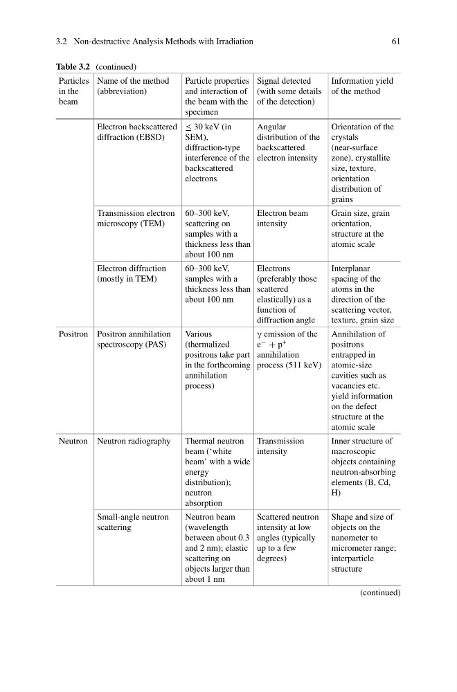
Table 3.2 (continued)
Particles
in the
beam
Name of the method
(abbreviation)
Particle properties
and interaction of
the beam with the
specimen
Signal detected
(with some details
of the detection)
Information yield
of the method
Electron backscattered
diffraction (EBSD)
≤ 30 keV (in
SEM),
diffraction-type
interference of the
backscattered
electrons
Angular
distribution of the
backscattered
electron intensity
Orientation of the
crystals
(near-surface
zone), crystallite
size, texture,
orientation
distribution of
grains
Transmission electron
microscopy (TEM)
60–300 keV,
scattering on
samples with a
thickness less than
about 100 nm
Electron beam
intensity
Grain size, grain
orientation,
structure at the
atomic scale
Electron diffraction
(mostly in TEM)
60–300 keV,
samples with a
thickness less than
about 100 nm
Electrons
(preferably those
scattered
elastically) as a
function of
diffraction angle
Interplanar
spacing of the
atoms in the
direction of the
scattering vector,
texture, grain size
Positron Positron annihilation
spectroscopy (PAS)
Various
(thermalized
positrons take part
in the forthcoming
annihilation
process)
γ emission of the
e − + p +
annihilation
process (511 keV)
Annihilation of
positrons
entrapped in
atomic-size
cavities such as
vacancies etc.
yield information
on the defect
structure at the
atomic scale
Neutron Neutron radiography
Thermal neutron
beam (‘white
beam’ with a wide
energy
distribution);
neutron
absorption
Transmission
intensity
Inner structure of
macroscopic
objects containing
neutron-absorbing
elements (B, Cd,
H)
Small-angle neutron
scattering
Neutron beam
(wavelength
between about 0.3
and 2 nm); elastic
scattering on
objects larger than
about 1 nm
Scattered neutron
intensity at low
angles (typically
up to a few
degrees)
Shape and size of
objects on the
nanometer to
micrometer range;
interparticle
structure
(continued)
61
Table 3.2 (continued)
Particles
in the
beam
Name of the method
(abbreviation)
Particle properties
and interaction of
the beam with the
specimen
Signal detected
(with some details
of the detection)
Information yield
of the method
Electron backscattered
diffraction (EBSD)
≤ 30 keV (in
SEM),
diffraction-type
interference of the
backscattered
electrons
Angular
distribution of the
backscattered
electron intensity
Orientation of the
crystals
(near-surface
zone), crystallite
size, texture,
orientation
distribution of
grains
Transmission electron
microscopy (TEM)
60–300 keV,
scattering on
samples with a
thickness less than
about 100 nm
Electron beam
intensity
Grain size, grain
orientation,
structure at the
atomic scale
Electron diffraction
(mostly in TEM)
60–300 keV,
samples with a
thickness less than
about 100 nm
Electrons
(preferably those
scattered
elastically) as a
function of
diffraction angle
Interplanar
spacing of the
atoms in the
direction of the
scattering vector,
texture, grain size
Positron Positron annihilation
spectroscopy (PAS)
Various
(thermalized
positrons take part
in the forthcoming
annihilation
process)
γ emission of the
e − + p +
annihilation
process (511 keV)
Annihilation of
positrons
entrapped in
atomic-size
cavities such as
vacancies etc.
yield information
on the defect
structure at the
atomic scale
Neutron Neutron radiography
Thermal neutron
beam (‘white
beam’ with a wide
energy
distribution);
neutron
absorption
Transmission
intensity
Inner structure of
macroscopic
objects containing
neutron-absorbing
elements (B, Cd,
H)
Small-angle neutron
scattering
Neutron beam
(wavelength
between about 0.3
and 2 nm); elastic
scattering on
objects larger than
about 1 nm
Scattered neutron
intensity at low
angles (typically
up to a few
degrees)
Shape and size of
objects on the
nanometer to
micrometer range;
interparticle
structure
(continued)