

60
3 Experimental Methods in Characterization of Nanosystems
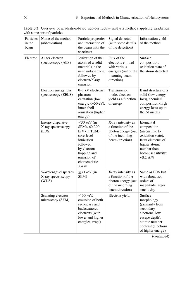
Table 3.2 Overview of irradiation-based non-destructive analysis methods applying irradiation
with some sort of particles
Particles
in the
beam
Name of the method
(abbreviation)
Particle properties
and interaction of
the beam with the
specimen
Signal detected
(with some details
of the detection)
Information yield
of the method
Electron Auger electron
spectroscopy (AES)
Ionization of the
atoms of a solid
material (in the
near surface zone)
followed by
electron/X-ray
emission
Flux of the
electrons emitted
with various
energies (out of the
incoming beam
direction)
Surface
composition,
oxidation state of
the atoms detected
Electron energy loss
spectroscopy (EELS)
0–1 kV electrons:
plasmon
excitation (low
energy, <~50 eV),
inner-shell
ionization (higher
energy)
Transmission
mode, electron
yield as a function
of energy
Band structure of a
solid (low energy
loss), chemical
composition (high
energy loss) up to
the 3d metals
Energy dispersive
X-ray spectroscopy
(EDS)
≤30 keV (in
SEM), 60-300
keV (in TEM);
core-level
ionization
followed
by electron
hopping and
emission of
characteristic
X-ray
X-ray intensity as
a function of the
photon energy (out
of the incoming
beam direction)
Elemental
composition
(insensitive to
oxidation state),
from elements of
higher atomic
number than
boron; sensitivity:
~0.2 at.%
Wavelength-dispersive
X-ray spectroscopy
(WDS)
≤30 keV (in
SEM)
X-ray intensity as
a function of the
photon energy (out
of the incoming
beam direction)
Same as EDS but
with about two
orders of
magnitude larger
sensitivity
Scanning electron
microscopy (SEM)
≤ 30 keV,
emission of both
secondary and
backscattered
electrons (with
lower and higher
energies, resp.)
Electron yield
Surface
morphology
(primarily from
secondary
electrons, low
escape depth),
atomic number
contrast (electrons
of higher energy)
(continued)
3 Experimental Methods in Characterization of Nanosystems
Table 3.2 Overview of irradiation-based non-destructive analysis methods applying irradiation
with some sort of particles
Particles
in the
beam
Name of the method
(abbreviation)
Particle properties
and interaction of
the beam with the
specimen
Signal detected
(with some details
of the detection)
Information yield
of the method
Electron Auger electron
spectroscopy (AES)
Ionization of the
atoms of a solid
material (in the
near surface zone)
followed by
electron/X-ray
emission
Flux of the
electrons emitted
with various
energies (out of the
incoming beam
direction)
Surface
composition,
oxidation state of
the atoms detected
Electron energy loss
spectroscopy (EELS)
0–1 kV electrons:
plasmon
excitation (low
energy, <~50 eV),
inner-shell
ionization (higher
energy)
Transmission
mode, electron
yield as a function
of energy
Band structure of a
solid (low energy
loss), chemical
composition (high
energy loss) up to
the 3d metals
Energy dispersive
X-ray spectroscopy
(EDS)
≤30 keV (in
SEM), 60-300
keV (in TEM);
core-level
ionization
followed
by electron
hopping and
emission of
characteristic
X-ray
X-ray intensity as
a function of the
photon energy (out
of the incoming
beam direction)
Elemental
composition
(insensitive to
oxidation state),
from elements of
higher atomic
number than
boron; sensitivity:
~0.2 at.%
Wavelength-dispersive
X-ray spectroscopy
(WDS)
≤30 keV (in
SEM)
X-ray intensity as
a function of the
photon energy (out
of the incoming
beam direction)
Same as EDS but
with about two
orders of
magnitude larger
sensitivity
Scanning electron
microscopy (SEM)
≤ 30 keV,
emission of both
secondary and
backscattered
electrons (with
lower and higher
energies, resp.)
Electron yield
Surface
morphology
(primarily from
secondary
electrons, low
escape depth),
atomic number
contrast (electrons
of higher energy)
(continued)