

213
ual TVMV switches, as assessed in Subheading 3.4.1. For
instance, for fully active TVMV switches, 100 nM is suitable
while for largely predominantly autoinhibited TVMV switches,
10 μM is required to reliably measure the initial rate.
6. Monitor the reaction progress using a fluorescence 96-wellplate
reader
by
monitoring
the
release
of
7- methoxycoumarinyl-4-acetyl from the quenched TVMV
peptide substrate with ʎ ex/em of 330 and 405 nm. Monitoring
the reaction progress over the course of 90 min is usually sufficient to reliably extract data for quantitative analysis.
7. Extract initial rates from the change in fluorescence (after subtracting the background fluorescence in the no enzyme control
at each substrate concentration) and plot against the substrate
concentration.
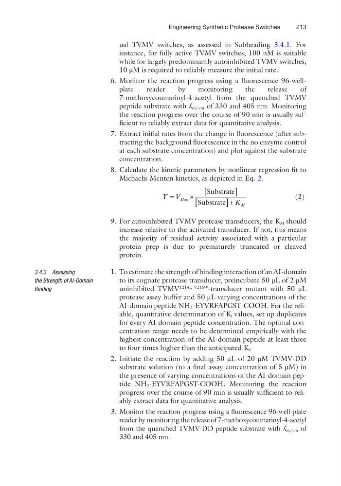
8. Calculate the kinetic parameters by nonlinear regression fit to
Michaelis Menten kinetics, as depicted in Eq. 2.
Y V
K
Max
M
=
´
[
]
[
]+
Substrate
Substrate
(2)
9. For autoinhibited TVMV protease transducers, the K M should
increase relative to the activated transducer. If not, this means
the majority of residual activity associated with a particular
protein prep is due to prematurely truncated or cleaved
protein.
1. To estimate the strength of binding interaction of an AI-domain
to its cognate protease transducer, preincubate 50 μL of 2 μM
uninhibited TVMV
T214I, V216W
-transducer mutant with 50 μL
protease assay buffer and 50 μL varying concentrations of the
AI-domain peptide NH 2 -EYVRFAPGST-COOH. For the reliable, quantitative determination of K i values, set up duplicates
for every AI-domain peptide concentration. The optimal concentration range needs to be determined empirically with the
highest concentration of the AI-domain peptide at least three
to four times higher than the anticipated K i .
2. Initiate the reaction by adding 50 μL of 20 μM TVMV-DD
substrate solution (to a final assay concentration of 5 μM) in
the presence of varying concentrations of the AI-domain peptide NH 2 -EYVRFAPGST-COOH. Monitoring the reaction
progress over the course of 90 min is usually sufficient to reliably extract data for quantitative analysis.
3. Monitor the reaction progress using a fluorescence 96-well- plate
reader by monitoring the release of 7- methoxycoumarinyl-4-acetyl
from the quenched TVMV-DD peptide substrate with ʎ ex/em of
330 and 405 nm.
3.4.3 Assessing
the Strength of AI-Domain
Binding
Engineering Synthetic Protease Switches
ual TVMV switches, as assessed in Subheading 3.4.1. For
instance, for fully active TVMV switches, 100 nM is suitable
while for largely predominantly autoinhibited TVMV switches,
10 μM is required to reliably measure the initial rate.
6. Monitor the reaction progress using a fluorescence 96-wellplate
reader
by
monitoring
the
release
of
7- methoxycoumarinyl-4-acetyl from the quenched TVMV
peptide substrate with ʎ ex/em of 330 and 405 nm. Monitoring
the reaction progress over the course of 90 min is usually sufficient to reliably extract data for quantitative analysis.
7. Extract initial rates from the change in fluorescence (after subtracting the background fluorescence in the no enzyme control
at each substrate concentration) and plot against the substrate
concentration.
8. Calculate the kinetic parameters by nonlinear regression fit to
Michaelis Menten kinetics, as depicted in Eq. 2.
Y V
K
Max
M
=
´
[
]
[
]+
Substrate
Substrate
(2)
9. For autoinhibited TVMV protease transducers, the K M should
increase relative to the activated transducer. If not, this means
the majority of residual activity associated with a particular
protein prep is due to prematurely truncated or cleaved
protein.
1. To estimate the strength of binding interaction of an AI-domain
to its cognate protease transducer, preincubate 50 μL of 2 μM
uninhibited TVMV
T214I, V216W
-transducer mutant with 50 μL
protease assay buffer and 50 μL varying concentrations of the
AI-domain peptide NH 2 -EYVRFAPGST-COOH. For the reliable, quantitative determination of K i values, set up duplicates
for every AI-domain peptide concentration. The optimal concentration range needs to be determined empirically with the
highest concentration of the AI-domain peptide at least three
to four times higher than the anticipated K i .
2. Initiate the reaction by adding 50 μL of 20 μM TVMV-DD
substrate solution (to a final assay concentration of 5 μM) in
the presence of varying concentrations of the AI-domain peptide NH 2 -EYVRFAPGST-COOH. Monitoring the reaction
progress over the course of 90 min is usually sufficient to reliably extract data for quantitative analysis.
3. Monitor the reaction progress using a fluorescence 96-well- plate
reader by monitoring the release of 7- methoxycoumarinyl-4-acetyl
from the quenched TVMV-DD peptide substrate with ʎ ex/em of
330 and 405 nm.
3.4.3 Assessing
the Strength of AI-Domain
Binding
Engineering Synthetic Protease Switches