

108
The distance D2 between the RET donor and RET acceptor in
the open state of the sensor can be controlled by modifying the
linker L2 which connects the SNAP-tag and the RET donor. Since
simple GGS linkers are conformationally flexible, they are not optimal as spacers [23]. Instead, polyproline linkers can be used, as
they form rigid helical structures [24]. In our experience, a
30- proline linker is sufficient to reduce RET efficiency in the open
state [25].
A list of all SNIFIT and LUCID sensors developed to date is
given (Table 1) while the following protocol provides a step-bystep summary how to construct and apply individual sensors.
2 Materials
1. RosettaGami DE3 pLysS electrocompetent E. coli cell.
2. LB medium: 5 g/L NaCl, 10 g/L tryptone, 5 g/L yeast
extract pH 7.5.
3. 100
mg/mL
ampicillin
stock
solution
(1000×),
filter-sterilized.
4. LB-agar plates supplemented with 100 μg/mL ampicillin.
5. 1 M isopropyl-β-thiogalactoside stock (IPTG) (1000×),
filter-sterilized.
6. Sonicator or French press.
2.1 Bacterial
Expression
and Purification
of SNIFITs and LUCIDs
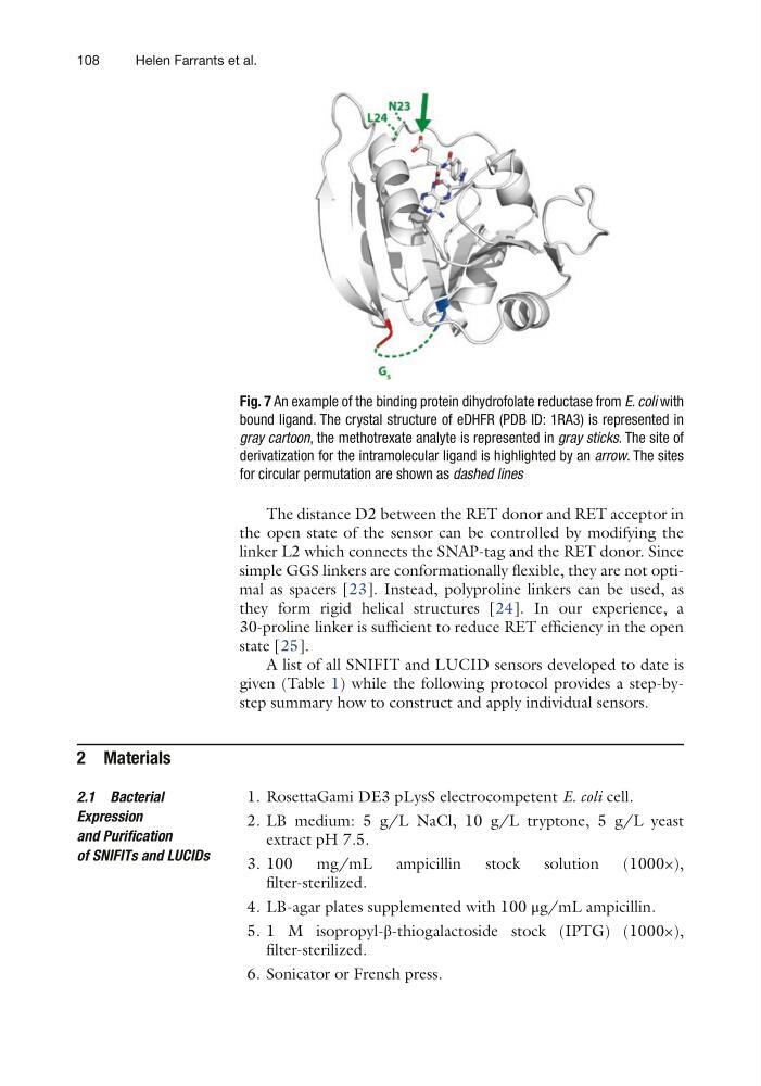
Fig. 7 An example of the binding protein dihydrofolate reductase from E. coli with
bound ligand. The crystal structure of eDHFR (PDB ID: 1RA3) is represented in
gray cartoon, the methotrexate analyte is represented in gray sticks. The site of
derivatization for the intramolecular ligand is highlighted by an arrow. The sites
for circular permutation are shown as dashed lines
Helen Farrants et al.
The distance D2 between the RET donor and RET acceptor in
the open state of the sensor can be controlled by modifying the
linker L2 which connects the SNAP-tag and the RET donor. Since
simple GGS linkers are conformationally flexible, they are not optimal as spacers [23]. Instead, polyproline linkers can be used, as
they form rigid helical structures [24]. In our experience, a
30- proline linker is sufficient to reduce RET efficiency in the open
state [25].
A list of all SNIFIT and LUCID sensors developed to date is
given (Table 1) while the following protocol provides a step-bystep summary how to construct and apply individual sensors.
2 Materials
1. RosettaGami DE3 pLysS electrocompetent E. coli cell.
2. LB medium: 5 g/L NaCl, 10 g/L tryptone, 5 g/L yeast
extract pH 7.5.
3. 100
mg/mL
ampicillin
stock
solution
(1000×),
filter-sterilized.
4. LB-agar plates supplemented with 100 μg/mL ampicillin.
5. 1 M isopropyl-β-thiogalactoside stock (IPTG) (1000×),
filter-sterilized.
6. Sonicator or French press.
2.1 Bacterial
Expression
and Purification
of SNIFITs and LUCIDs
Fig. 7 An example of the binding protein dihydrofolate reductase from E. coli with
bound ligand. The crystal structure of eDHFR (PDB ID: 1RA3) is represented in
gray cartoon, the methotrexate analyte is represented in gray sticks. The site of
derivatization for the intramolecular ligand is highlighted by an arrow. The sites
for circular permutation are shown as dashed lines
Helen Farrants et al.