

414
I. de Moraes and N. M. Dempsey
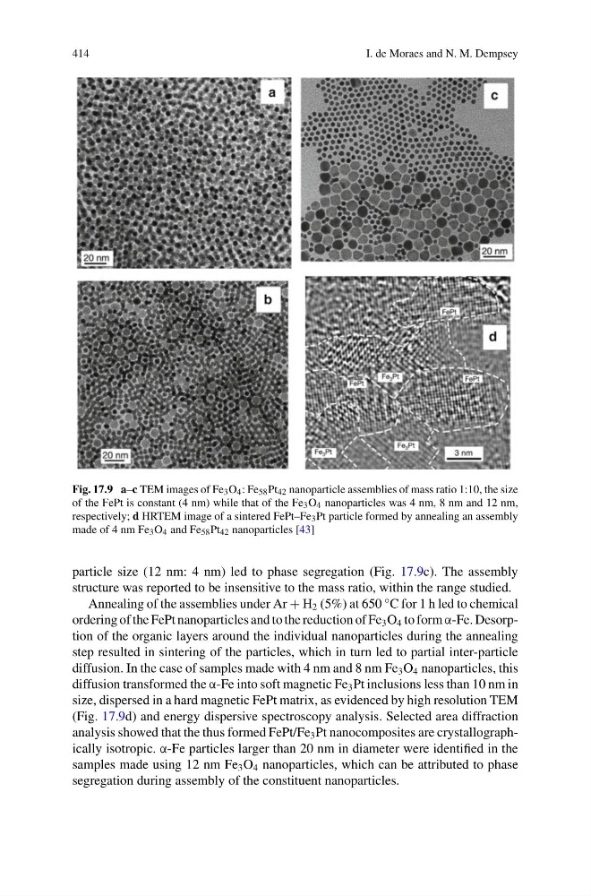
Fig. 17.9 a–c TEM images of Fe 3 O 4 : Fe 58 Pt 42 nanoparticle assemblies of mass ratio 1:10, the size
of the FePt is constant (4 nm) while that of the Fe 3 O 4 nanoparticles was 4 nm, 8 nm and 12 nm,
respectively; d HRTEM image of a sintered FePt–Fe 3 Pt particle formed by annealing an assembly
made of 4 nm Fe 3 O 4 and Fe 58 Pt 42 nanoparticles [43]
particle size (12 nm: 4 nm) led to phase segregation (Fig. 17.9c). The assembly
structure was reported to be insensitive to the mass ratio, within the range studied.
Annealing of the assemblies under Ar + H 2 (5%) at 650 °C for 1 h led to chemical
ordering of the FePt nanoparticles and to the reduction of Fe 3 O 4 to form α-Fe. Desorption of the organic layers around the individual nanoparticles during the annealing
step resulted in sintering of the particles, which in turn led to partial inter-particle
diffusion. In the case of samples made with 4 nm and 8 nm Fe 3 O 4 nanoparticles, this
diffusion transformed the α-Fe into soft magnetic Fe 3 Pt inclusions less than 10 nm in
size, dispersed in a hard magnetic FePt matrix, as evidenced by high resolution TEM
(Fig. 17.9d) and energy dispersive spectroscopy analysis. Selected area diffraction
analysis showed that the thus formed FePt/Fe 3 Pt nanocomposites are crystallographically isotropic. α-Fe particles larger than 20 nm in diameter were identified in the
samples made using 12 nm Fe 3 O 4 nanoparticles, which can be attributed to phase
segregation during assembly of the constituent nanoparticles.
I. de Moraes and N. M. Dempsey
Fig. 17.9 a–c TEM images of Fe 3 O 4 : Fe 58 Pt 42 nanoparticle assemblies of mass ratio 1:10, the size
of the FePt is constant (4 nm) while that of the Fe 3 O 4 nanoparticles was 4 nm, 8 nm and 12 nm,
respectively; d HRTEM image of a sintered FePt–Fe 3 Pt particle formed by annealing an assembly
made of 4 nm Fe 3 O 4 and Fe 58 Pt 42 nanoparticles [43]
particle size (12 nm: 4 nm) led to phase segregation (Fig. 17.9c). The assembly
structure was reported to be insensitive to the mass ratio, within the range studied.
Annealing of the assemblies under Ar + H 2 (5%) at 650 °C for 1 h led to chemical
ordering of the FePt nanoparticles and to the reduction of Fe 3 O 4 to form α-Fe. Desorption of the organic layers around the individual nanoparticles during the annealing
step resulted in sintering of the particles, which in turn led to partial inter-particle
diffusion. In the case of samples made with 4 nm and 8 nm Fe 3 O 4 nanoparticles, this
diffusion transformed the α-Fe into soft magnetic Fe 3 Pt inclusions less than 10 nm in
size, dispersed in a hard magnetic FePt matrix, as evidenced by high resolution TEM
(Fig. 17.9d) and energy dispersive spectroscopy analysis. Selected area diffraction
analysis showed that the thus formed FePt/Fe 3 Pt nanocomposites are crystallographically isotropic. α-Fe particles larger than 20 nm in diameter were identified in the
samples made using 12 nm Fe 3 O 4 nanoparticles, which can be attributed to phase
segregation during assembly of the constituent nanoparticles.