

13 Magnetic Nanoparticles for Life Sciences Applications
319
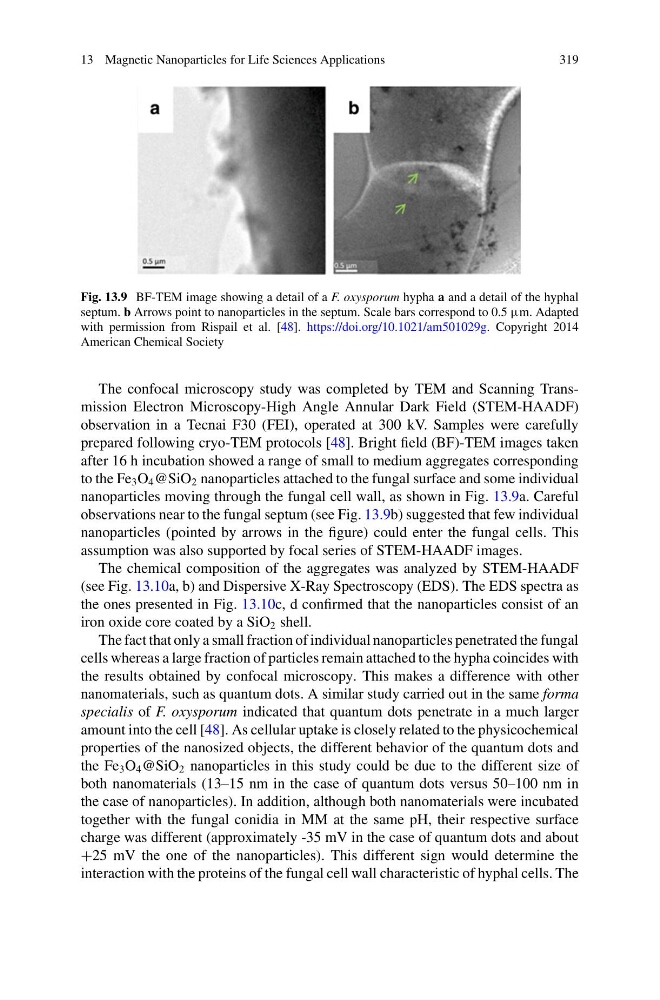
Fig. 13.9 BF-TEM image showing a detail of a F. oxysporum hypha a and a detail of the hyphal
septum. b Arrows point to nanoparticles in the septum. Scale bars correspond to 0.5 μm. Adapted
with permission from Rispail et al. [48]. https://doi.org/10.1021/am501029g. Copyright 2014
American Chemical Society
The confocal microscopy study was completed by TEM and Scanning Transmission Electron Microscopy-High Angle Annular Dark Field (STEM-HAADF)
observation in a Tecnai F30 (FEI), operated at 300 kV. Samples were carefully
prepared following cryo-TEM protocols [48]. Bright field (BF)-TEM images taken
after 16 h incubation showed a range of small to medium aggregates corresponding
to the Fe 3 O 4 @SiO 2 nanoparticles attached to the fungal surface and some individual
nanoparticles moving through the fungal cell wall, as shown in Fig. 13.9a. Careful
observations near to the fungal septum (see Fig. 13.9b) suggested that few individual
nanoparticles (pointed by arrows in the figure) could enter the fungal cells. This
assumption was also supported by focal series of STEM-HAADF images.
The chemical composition of the aggregates was analyzed by STEM-HAADF
(see Fig. 13.10a, b) and Dispersive X-Ray Spectroscopy (EDS). The EDS spectra as
the ones presented in Fig. 13.10c, d confirmed that the nanoparticles consist of an
iron oxide core coated by a SiO 2 shell.
The fact that only a small fraction of individual nanoparticles penetrated the fungal
cells whereas a large fraction of particles remain attached to the hypha coincides with
the results obtained by confocal microscopy. This makes a difference with other
nanomaterials, such as quantum dots. A similar study carried out in the same forma
specialis of F. oxysporum indicated that quantum dots penetrate in a much larger
amount into the cell [48]. As cellular uptake is closely related to the physicochemical
properties of the nanosized objects, the different behavior of the quantum dots and
the Fe 3 O 4 @SiO 2 nanoparticles in this study could be due to the different size of
both nanomaterials (13–15 nm in the case of quantum dots versus 50–100 nm in
the case of nanoparticles). In addition, although both nanomaterials were incubated
together with the fungal conidia in MM at the same pH, their respective surface
charge was different (approximately -35 mV in the case of quantum dots and about
+25 mV the one of the nanoparticles). This different sign would determine the
interaction with the proteins of the fungal cell wall characteristic of hyphal cells. The
319
Fig. 13.9 BF-TEM image showing a detail of a F. oxysporum hypha a and a detail of the hyphal
septum. b Arrows point to nanoparticles in the septum. Scale bars correspond to 0.5 μm. Adapted
with permission from Rispail et al. [48]. https://doi.org/10.1021/am501029g. Copyright 2014
American Chemical Society
The confocal microscopy study was completed by TEM and Scanning Transmission Electron Microscopy-High Angle Annular Dark Field (STEM-HAADF)
observation in a Tecnai F30 (FEI), operated at 300 kV. Samples were carefully
prepared following cryo-TEM protocols [48]. Bright field (BF)-TEM images taken
after 16 h incubation showed a range of small to medium aggregates corresponding
to the Fe 3 O 4 @SiO 2 nanoparticles attached to the fungal surface and some individual
nanoparticles moving through the fungal cell wall, as shown in Fig. 13.9a. Careful
observations near to the fungal septum (see Fig. 13.9b) suggested that few individual
nanoparticles (pointed by arrows in the figure) could enter the fungal cells. This
assumption was also supported by focal series of STEM-HAADF images.
The chemical composition of the aggregates was analyzed by STEM-HAADF
(see Fig. 13.10a, b) and Dispersive X-Ray Spectroscopy (EDS). The EDS spectra as
the ones presented in Fig. 13.10c, d confirmed that the nanoparticles consist of an
iron oxide core coated by a SiO 2 shell.
The fact that only a small fraction of individual nanoparticles penetrated the fungal
cells whereas a large fraction of particles remain attached to the hypha coincides with
the results obtained by confocal microscopy. This makes a difference with other
nanomaterials, such as quantum dots. A similar study carried out in the same forma
specialis of F. oxysporum indicated that quantum dots penetrate in a much larger
amount into the cell [48]. As cellular uptake is closely related to the physicochemical
properties of the nanosized objects, the different behavior of the quantum dots and
the Fe 3 O 4 @SiO 2 nanoparticles in this study could be due to the different size of
both nanomaterials (13–15 nm in the case of quantum dots versus 50–100 nm in
the case of nanoparticles). In addition, although both nanomaterials were incubated
together with the fungal conidia in MM at the same pH, their respective surface
charge was different (approximately -35 mV in the case of quantum dots and about
+25 mV the one of the nanoparticles). This different sign would determine the
interaction with the proteins of the fungal cell wall characteristic of hyphal cells. The