

318
C. Marquina
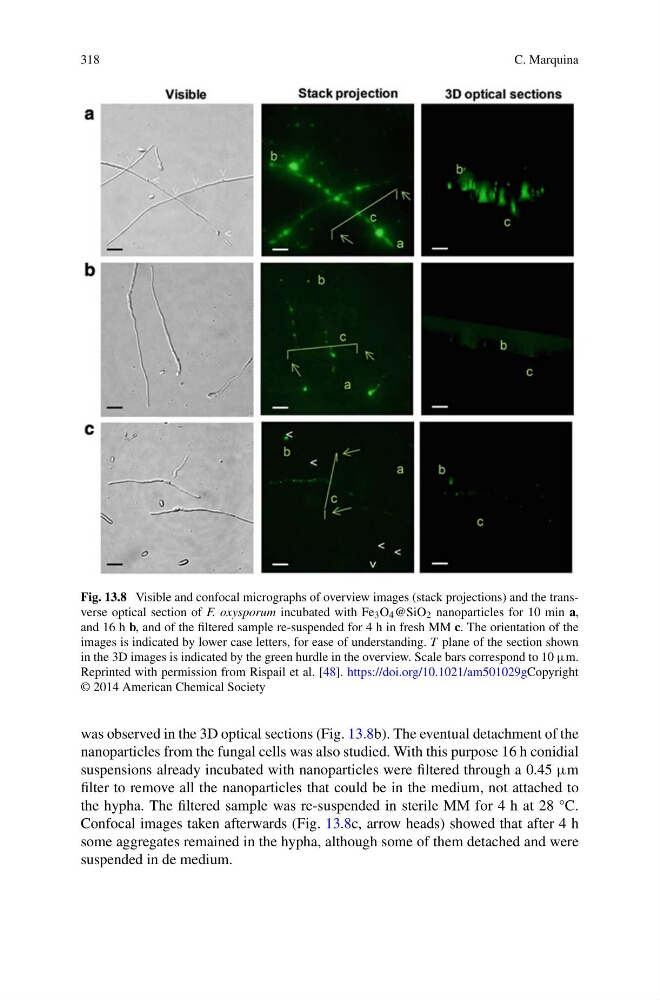
Fig. 13.8 Visible and confocal micrographs of overview images (stack projections) and the transverse optical section of F. oxysporum incubated with Fe 3 O 4 @SiO 2 nanoparticles for 10 min a,
and 16 h b, and of the filtered sample re-suspended for 4 h in fresh MM c. The orientation of the
images is indicated by lower case letters, for ease of understanding. T plane of the section shown
in the 3D images is indicated by the green hurdle in the overview. Scale bars correspond to 10 μm.
Reprinted with permission from Rispail et al. [48]. https://doi.org/10.1021/am501029gCopyright
© 2014 American Chemical Society
was observed in the 3D optical sections (Fig. 13.8b). The eventual detachment of the
nanoparticles from the fungal cells was also studied. With this purpose 16 h conidial
suspensions already incubated with nanoparticles were filtered through a 0.45 μm
filter to remove all the nanoparticles that could be in the medium, not attached to
the hypha. The filtered sample was re-suspended in sterile MM for 4 h at 28 °C.
Confocal images taken afterwards (Fig. 13.8c, arrow heads) showed that after 4 h
some aggregates remained in the hypha, although some of them detached and were
suspended in de medium.
C. Marquina
Fig. 13.8 Visible and confocal micrographs of overview images (stack projections) and the transverse optical section of F. oxysporum incubated with Fe 3 O 4 @SiO 2 nanoparticles for 10 min a,
and 16 h b, and of the filtered sample re-suspended for 4 h in fresh MM c. The orientation of the
images is indicated by lower case letters, for ease of understanding. T plane of the section shown
in the 3D images is indicated by the green hurdle in the overview. Scale bars correspond to 10 μm.
Reprinted with permission from Rispail et al. [48]. https://doi.org/10.1021/am501029gCopyright
© 2014 American Chemical Society
was observed in the 3D optical sections (Fig. 13.8b). The eventual detachment of the
nanoparticles from the fungal cells was also studied. With this purpose 16 h conidial
suspensions already incubated with nanoparticles were filtered through a 0.45 μm
filter to remove all the nanoparticles that could be in the medium, not attached to
the hypha. The filtered sample was re-suspended in sterile MM for 4 h at 28 °C.
Confocal images taken afterwards (Fig. 13.8c, arrow heads) showed that after 4 h
some aggregates remained in the hypha, although some of them detached and were
suspended in de medium.