

308
C. Marquina
13.2.1 Nanoparticle Application by Injection
These experiments were carried out on living pumpkin plants and with the Fe@C
nanoparticles suspended in gelafundine. The pumpkin plants were selected because
of the large size of their vessels, to make the transport of the nanoparticles through
the vascular system easier. Plants were grown as described in [52, 54, 63]. In a
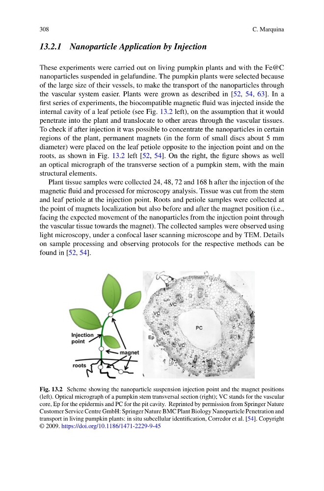
first series of experiments, the biocompatible magnetic fluid was injected inside the
internal cavity of a leaf petiole (see Fig. 13.2 left), on the assumption that it would
penetrate into the plant and translocate to other areas through the vascular tissues.
To check if after injection it was possible to concentrate the nanoparticles in certain
regions of the plant, permanent magnets (in the form of small discs about 5 mm
diameter) were placed on the leaf petiole opposite to the injection point and on the
roots, as shown in Fig. 13.2 left [52, 54]. On the right, the figure shows as well
an optical micrograph of the transverse section of a pumpkin stem, with the main
structural elements.
Plant tissue samples were collected 24, 48, 72 and 168 h after the injection of the
magnetic fluid and processed for microscopy analysis. Tissue was cut from the stem
and leaf petiole at the injection point. Roots and petiole samples were collected at
the point of magnets localization but also before and after the magnet position (i.e.,
facing the expected movement of the nanoparticles from the injection point through
the vascular tissue towards the magnet). The collected samples were observed using
light microscopy, under a confocal laser scanning microscope and by TEM. Details
on sample processing and observing protocols for the respective methods can be
found in [52, 54].
Fig. 13.2 Scheme showing the nanoparticle suspension injection point and the magnet positions
(left). Optical micrograph of a pumpkin stem transversal section (right); VC stands for the vascular
core, Ep for the epidermis and PC for the pit cavity. Reprinted by permission from Springer Nature
Customer Service Centre GmbH: Springer Nature BMC Plant Biology Nanoparticle Penetration and
transport in living pumpkin plants: in situ subcellular identification, Corredor et al. [54]. Copyright
© 2009. https://doi.org/10.1186/1471-2229-9-45
C. Marquina
13.2.1 Nanoparticle Application by Injection
These experiments were carried out on living pumpkin plants and with the Fe@C
nanoparticles suspended in gelafundine. The pumpkin plants were selected because
of the large size of their vessels, to make the transport of the nanoparticles through
the vascular system easier. Plants were grown as described in [52, 54, 63]. In a
first series of experiments, the biocompatible magnetic fluid was injected inside the
internal cavity of a leaf petiole (see Fig. 13.2 left), on the assumption that it would
penetrate into the plant and translocate to other areas through the vascular tissues.
To check if after injection it was possible to concentrate the nanoparticles in certain
regions of the plant, permanent magnets (in the form of small discs about 5 mm
diameter) were placed on the leaf petiole opposite to the injection point and on the
roots, as shown in Fig. 13.2 left [52, 54]. On the right, the figure shows as well
an optical micrograph of the transverse section of a pumpkin stem, with the main
structural elements.
Plant tissue samples were collected 24, 48, 72 and 168 h after the injection of the
magnetic fluid and processed for microscopy analysis. Tissue was cut from the stem
and leaf petiole at the injection point. Roots and petiole samples were collected at
the point of magnets localization but also before and after the magnet position (i.e.,
facing the expected movement of the nanoparticles from the injection point through
the vascular tissue towards the magnet). The collected samples were observed using
light microscopy, under a confocal laser scanning microscope and by TEM. Details
on sample processing and observing protocols for the respective methods can be
found in [52, 54].
Fig. 13.2 Scheme showing the nanoparticle suspension injection point and the magnet positions
(left). Optical micrograph of a pumpkin stem transversal section (right); VC stands for the vascular
core, Ep for the epidermis and PC for the pit cavity. Reprinted by permission from Springer Nature
Customer Service Centre GmbH: Springer Nature BMC Plant Biology Nanoparticle Penetration and
transport in living pumpkin plants: in situ subcellular identification, Corredor et al. [54]. Copyright
© 2009. https://doi.org/10.1186/1471-2229-9-45