

13 Magnetic Nanoparticles for Life Sciences Applications
307
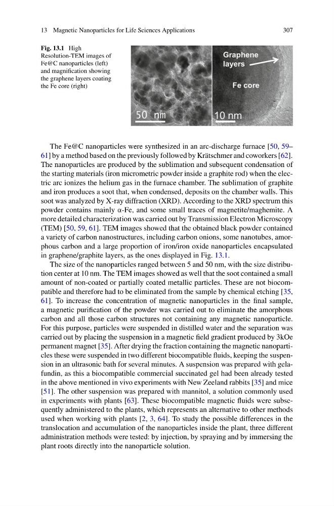
Fig. 13.1 High
Resolution-TEM images of
Fe@C nanoparticles (left)
and magnification showing
the graphene layers coating
the Fe core (right)
The Fe@C nanoparticles were synthesized in an arc-discharge furnace [50, 59–
61] by a method based on the previously followed by Krätschmer and coworkers [62].
The nanoparticles are produced by the sublimation and subsequent condensation of
the starting materials (iron micrometric powder inside a graphite rod) when the electric arc ionizes the helium gas in the furnace chamber. The sublimation of graphite
and iron produces a soot that, when condensed, deposits on the chamber walls. This
soot was analyzed by X-ray diffraction (XRD). According to the XRD spectrum this
powder contains mainly α-Fe, and some small traces of magnetite/maghemite. A
more detailed characterization was carried out by Transmission Electron Microscopy
(TEM) [50, 59, 61]. TEM images showed that the obtained black powder contained
a variety of carbon nanostructures, including carbon onions, some nanotubes, amorphous carbon and a large proportion of iron/iron oxide nanoparticles encapsulated
in graphene/graphite layers, as the ones displayed in Fig. 13.1.
The size of the nanoparticles ranged between 5 and 50 nm, with the size distribution center at 10 nm. The TEM images showed as well that the soot contained a small
amount of non-coated or partially coated metallic particles. These are not biocompatible and therefore had to be eliminated from the sample by chemical etching [35,
61]. To increase the concentration of magnetic nanoparticles in the final sample,
a magnetic purification of the powder was carried out to eliminate the amorphous
carbon and all those carbon structures not containing any magnetic nanoparticle.
For this purpose, particles were suspended in distilled water and the separation was
carried out by placing the suspension in a magnetic field gradient produced by 3kOe
permanent magnet [35]. After drying the fraction containing the magnetic nanoparticles these were suspended in two different biocompatible fluids, keeping the suspension in an ultrasonic bath for several minutes. A suspension was prepared with gelafundin, as this a biocompatible commercial succinated gel had been already tested
in the above mentioned in vivo experiments with New Zeeland rabbits [35] and mice
[51]. The other suspension was prepared with mannitol, a solution commonly used
in experiments with plants [63]. These biocompatible magnetic fluids were subsequently administered to the plants, which represents an alternative to other methods
used when working with plants [2, 3, 64]. To study the possible differences in the
translocation and accumulation of the nanoparticles inside the plant, three different
administration methods were tested: by injection, by spraying and by immersing the
plant roots directly into the nanoparticle solution.
307
Fig. 13.1 High
Resolution-TEM images of
Fe@C nanoparticles (left)
and magnification showing
the graphene layers coating
the Fe core (right)
The Fe@C nanoparticles were synthesized in an arc-discharge furnace [50, 59–
61] by a method based on the previously followed by Krätschmer and coworkers [62].
The nanoparticles are produced by the sublimation and subsequent condensation of
the starting materials (iron micrometric powder inside a graphite rod) when the electric arc ionizes the helium gas in the furnace chamber. The sublimation of graphite
and iron produces a soot that, when condensed, deposits on the chamber walls. This
soot was analyzed by X-ray diffraction (XRD). According to the XRD spectrum this
powder contains mainly α-Fe, and some small traces of magnetite/maghemite. A
more detailed characterization was carried out by Transmission Electron Microscopy
(TEM) [50, 59, 61]. TEM images showed that the obtained black powder contained
a variety of carbon nanostructures, including carbon onions, some nanotubes, amorphous carbon and a large proportion of iron/iron oxide nanoparticles encapsulated
in graphene/graphite layers, as the ones displayed in Fig. 13.1.
The size of the nanoparticles ranged between 5 and 50 nm, with the size distribution center at 10 nm. The TEM images showed as well that the soot contained a small
amount of non-coated or partially coated metallic particles. These are not biocompatible and therefore had to be eliminated from the sample by chemical etching [35,
61]. To increase the concentration of magnetic nanoparticles in the final sample,
a magnetic purification of the powder was carried out to eliminate the amorphous
carbon and all those carbon structures not containing any magnetic nanoparticle.
For this purpose, particles were suspended in distilled water and the separation was
carried out by placing the suspension in a magnetic field gradient produced by 3kOe
permanent magnet [35]. After drying the fraction containing the magnetic nanoparticles these were suspended in two different biocompatible fluids, keeping the suspension in an ultrasonic bath for several minutes. A suspension was prepared with gelafundin, as this a biocompatible commercial succinated gel had been already tested
in the above mentioned in vivo experiments with New Zeeland rabbits [35] and mice
[51]. The other suspension was prepared with mannitol, a solution commonly used
in experiments with plants [63]. These biocompatible magnetic fluids were subsequently administered to the plants, which represents an alternative to other methods
used when working with plants [2, 3, 64]. To study the possible differences in the
translocation and accumulation of the nanoparticles inside the plant, three different
administration methods were tested: by injection, by spraying and by immersing the
plant roots directly into the nanoparticle solution.