

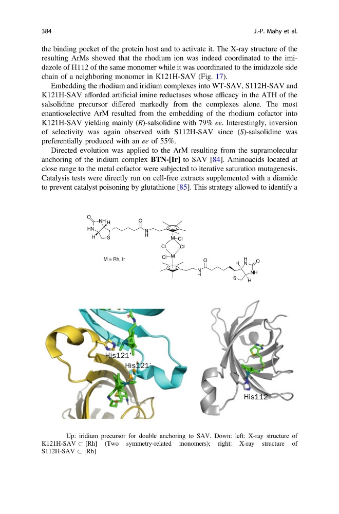
the binding pocket of the protein host and to activate it. The X-ray structure of the
resulting ArMs showed that the rhodium ion was indeed coordinated to the imidazole of H112 of the same monomer while it was coordinated to the imidazole side
chain of a neighboring monomer in K121H-SAV (Fig. 17).
Embedding the rhodium and iridium complexes into WT-SAV, S112H-SAV and
K121H-SAV afforded artificial imine reductases whose efficacy in the ATH of the
salsolidine precursor differed markedly from the complexes alone. The most
enantioselective ArM resulted from the embedding of the rhodium cofactor into
K121H-SAV yielding mainly (R)-salsolidine with 79% ee. Interestingly, inversion
of selectivity was again observed with S112H-SAV since (S)-salsolidine was
preferentially produced with an ee of 55%.
Directed evolution was applied to the ArM resulting from the supramolecular
anchoring of the iridium complex BTN-[Ir] to SAV [84]. Aminoacids located at
close range to the metal cofactor were subjected to iterative saturation mutagenesis.
Catalysis tests were directly run on cell-free extracts supplemented with a diamide
to prevent catalyst poisoning by glutathione [85]. This strategy allowed to identify a
His121’
His121’
His112
Fig. 17 Up: iridium precursor for double anchoring to SAV. Down: left: X-ray structure of
K121H-SAV & [Rh] (Two symmetry-related monomers); right: X-ray structure of
S112H-SAV & [Rh]
384
J.-P. Mahy et al.
resulting ArMs showed that the rhodium ion was indeed coordinated to the imidazole of H112 of the same monomer while it was coordinated to the imidazole side
chain of a neighboring monomer in K121H-SAV (Fig. 17).
Embedding the rhodium and iridium complexes into WT-SAV, S112H-SAV and
K121H-SAV afforded artificial imine reductases whose efficacy in the ATH of the
salsolidine precursor differed markedly from the complexes alone. The most
enantioselective ArM resulted from the embedding of the rhodium cofactor into
K121H-SAV yielding mainly (R)-salsolidine with 79% ee. Interestingly, inversion
of selectivity was again observed with S112H-SAV since (S)-salsolidine was
preferentially produced with an ee of 55%.
Directed evolution was applied to the ArM resulting from the supramolecular
anchoring of the iridium complex BTN-[Ir] to SAV [84]. Aminoacids located at
close range to the metal cofactor were subjected to iterative saturation mutagenesis.
Catalysis tests were directly run on cell-free extracts supplemented with a diamide
to prevent catalyst poisoning by glutathione [85]. This strategy allowed to identify a
His121’
His121’
His112
Fig. 17 Up: iridium precursor for double anchoring to SAV. Down: left: X-ray structure of
K121H-SAV & [Rh] (Two symmetry-related monomers); right: X-ray structure of
S112H-SAV & [Rh]
384
J.-P. Mahy et al.