

Another interesting doped system that has been studied is Eu
2þ doped in a ZnS
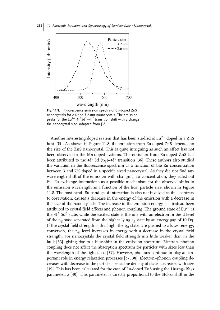
host [35]. As shown in Figure 11.8, the emission from Eu-doped ZnS depends on
the size of the ZnS nanocrystal. This is quite intriguing as such an effect has not
been observed in the Mn-doped systems. The emission from Eu-doped ZnS has
been attributed to the 4f
6 5d
1 (t 2g )–4f
7 transition [36]. These authors also studied
the variation in the fluorescence spectrum as a function of the Eu concentration
between 3 and 7% doped in a specific sized nanocrystal. As they did not find any
wavelength shift of the emission with changing Eu concentration, they ruled out
EuaEu exchange interactions as a possible mechanism for the observed shifts in
the emission wavelength as a function of the host particle size, shown in Figure
11.8. The host band–Eu band sp–d interaction is also not involved as this, contrary
to observation, causes a decrease in the energy of the emission with a decrease in
the size of the nanocrystals. The increase in the emission energy has instead been
attributed to crystal field effects and phonon coupling. The ground state of Eu
2þ is
the 4f
7 5d
0 state, while the excited state is the one with an electron in the d level
of the t 2g state separated from the higher lying e g state by an energy gap of 10 Dq.
If the crystal field strength is this high, the t 2g states are pushed to a lower energy;
conversely, the t 2g level increases in energy with a decrease in the crystal field
strength. For nanocrystals the crystal field strength is a little weaker than in the
bulk [33], giving rise to a blue-shift in the emission spectrum. Electron–phonon
coupling does not affect the absorption spectrum for particles with sizes less than
the wavelength of the light used [37]. However, phonons continue to play an important role in energy relaxation processes [37, 38]. Electron–phonon coupling decreases with decrease in the particle size as the density of states decreases with size
[39]. This has been calculated for the case of Eu-doped ZnS using the Huang–Rhys
parameter, S [40]. This parameter is directly proportional to the Stokes shift in the
400
500
600
700
Intensity (arb. units)
wavelength (nm)
Particle size
3.2 nm
2.6 nm
Fig. 11.8. Fluorescence emission spectra of Eu-doped ZnS
nanocrystals for 2.6 and 3.2 nm nanocrystals. The emission
peaks for the Eu
2þ 4f
6 5d
1 –4f
7 transition shift with a change in
the nanocrystal size. Adapted from [35].
11 Electronic Structure and Spectroscopy of Semiconductor Nanocrystals
382
2þ doped in a ZnS
host [35]. As shown in Figure 11.8, the emission from Eu-doped ZnS depends on
the size of the ZnS nanocrystal. This is quite intriguing as such an effect has not
been observed in the Mn-doped systems. The emission from Eu-doped ZnS has
been attributed to the 4f
6 5d
1 (t 2g )–4f
7 transition [36]. These authors also studied
the variation in the fluorescence spectrum as a function of the Eu concentration
between 3 and 7% doped in a specific sized nanocrystal. As they did not find any
wavelength shift of the emission with changing Eu concentration, they ruled out
EuaEu exchange interactions as a possible mechanism for the observed shifts in
the emission wavelength as a function of the host particle size, shown in Figure
11.8. The host band–Eu band sp–d interaction is also not involved as this, contrary
to observation, causes a decrease in the energy of the emission with a decrease in
the size of the nanocrystals. The increase in the emission energy has instead been
attributed to crystal field effects and phonon coupling. The ground state of Eu
2þ is
the 4f
7 5d
0 state, while the excited state is the one with an electron in the d level
of the t 2g state separated from the higher lying e g state by an energy gap of 10 Dq.
If the crystal field strength is this high, the t 2g states are pushed to a lower energy;
conversely, the t 2g level increases in energy with a decrease in the crystal field
strength. For nanocrystals the crystal field strength is a little weaker than in the
bulk [33], giving rise to a blue-shift in the emission spectrum. Electron–phonon
coupling does not affect the absorption spectrum for particles with sizes less than
the wavelength of the light used [37]. However, phonons continue to play an important role in energy relaxation processes [37, 38]. Electron–phonon coupling decreases with decrease in the particle size as the density of states decreases with size
[39]. This has been calculated for the case of Eu-doped ZnS using the Huang–Rhys
parameter, S [40]. This parameter is directly proportional to the Stokes shift in the
400
500
600
700
Intensity (arb. units)
wavelength (nm)
Particle size
3.2 nm
2.6 nm
Fig. 11.8. Fluorescence emission spectra of Eu-doped ZnS
nanocrystals for 2.6 and 3.2 nm nanocrystals. The emission
peaks for the Eu
2þ 4f
6 5d
1 –4f
7 transition shift with a change in
the nanocrystal size. Adapted from [35].
11 Electronic Structure and Spectroscopy of Semiconductor Nanocrystals
382