

11 Fluorescent Chemosensors
403
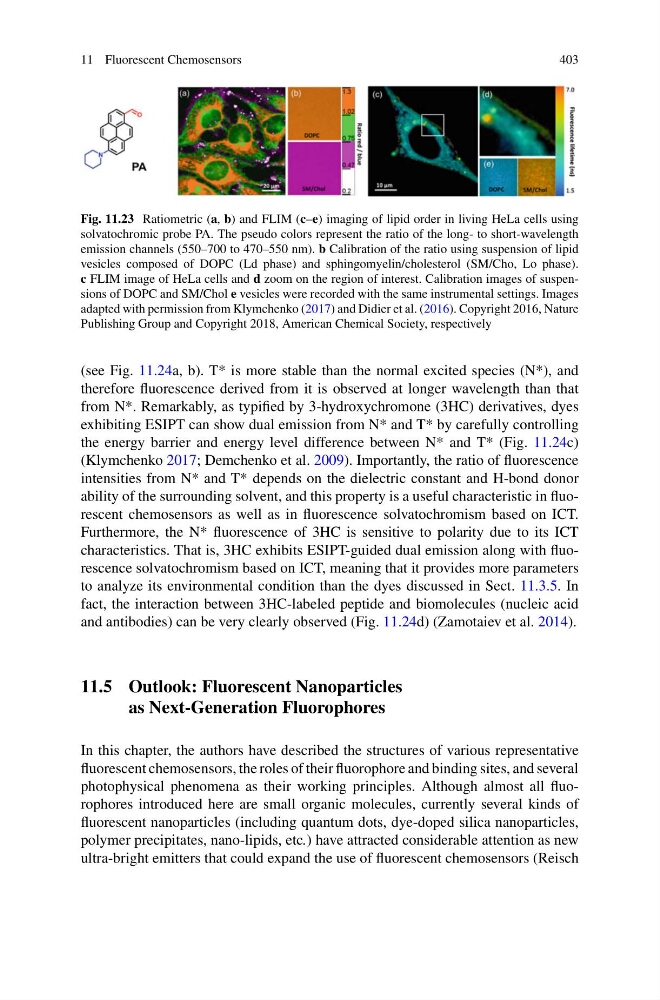
Fig. 11.23 Ratiometric (a, b) and FLIM (c–e) imaging of lipid order in living HeLa cells using
solvatochromic probe PA. The pseudo colors represent the ratio of the long- to short-wavelength
emission channels (550–700 to 470–550 nm). b Calibration of the ratio using suspension of lipid
vesicles composed of DOPC (Ld phase) and sphingomyelin/cholesterol (SM/Cho, Lo phase).
c FLIM image of HeLa cells and d zoom on the region of interest. Calibration images of suspensions of DOPC and SM/Chol e vesicles were recorded with the same instrumental settings. Images
adapted with permission from Klymchenko (2017) and Didier et al. (2016). Copyright 2016, Nature
Publishing Group and Copyright 2018, American Chemical Society, respectively
(see Fig. 11.24a, b). T* is more stable than the normal excited species (N*), and
therefore fluorescence derived from it is observed at longer wavelength than that
from N*. Remarkably, as typified by 3-hydroxychromone (3HC) derivatives, dyes
exhibiting ESIPT can show dual emission from N* and T* by carefully controlling
the energy barrier and energy level difference between N* and T* (Fig. 11.24c)
(Klymchenko 2017; Demchenko et al. 2009). Importantly, the ratio of fluorescence
intensities from N* and T* depends on the dielectric constant and H-bond donor
ability of the surrounding solvent, and this property is a useful characteristic in fluorescent chemosensors as well as in fluorescence solvatochromism based on ICT.
Furthermore, the N* fluorescence of 3HC is sensitive to polarity due to its ICT
characteristics. That is, 3HC exhibits ESIPT-guided dual emission along with fluorescence solvatochromism based on ICT, meaning that it provides more parameters
to analyze its environmental condition than the dyes discussed in Sect. 11.3.5. In
fact, the interaction between 3HC-labeled peptide and biomolecules (nucleic acid
and antibodies) can be very clearly observed (Fig. 11.24d) (Zamotaiev et al. 2014).
11.5 Outlook: Fluorescent Nanoparticles
as Next-Generation Fluorophores
In this chapter, the authors have described the structures of various representative
fluorescent chemosensors, the roles of their fluorophore and binding sites, and several
photophysical phenomena as their working principles. Although almost all fluorophores introduced here are small organic molecules, currently several kinds of
fluorescent nanoparticles (including quantum dots, dye-doped silica nanoparticles,
polymer precipitates, nano-lipids, etc.) have attracted considerable attention as new
ultra-bright emitters that could expand the use of fluorescent chemosensors (Reisch
403
Fig. 11.23 Ratiometric (a, b) and FLIM (c–e) imaging of lipid order in living HeLa cells using
solvatochromic probe PA. The pseudo colors represent the ratio of the long- to short-wavelength
emission channels (550–700 to 470–550 nm). b Calibration of the ratio using suspension of lipid
vesicles composed of DOPC (Ld phase) and sphingomyelin/cholesterol (SM/Cho, Lo phase).
c FLIM image of HeLa cells and d zoom on the region of interest. Calibration images of suspensions of DOPC and SM/Chol e vesicles were recorded with the same instrumental settings. Images
adapted with permission from Klymchenko (2017) and Didier et al. (2016). Copyright 2016, Nature
Publishing Group and Copyright 2018, American Chemical Society, respectively
(see Fig. 11.24a, b). T* is more stable than the normal excited species (N*), and
therefore fluorescence derived from it is observed at longer wavelength than that
from N*. Remarkably, as typified by 3-hydroxychromone (3HC) derivatives, dyes
exhibiting ESIPT can show dual emission from N* and T* by carefully controlling
the energy barrier and energy level difference between N* and T* (Fig. 11.24c)
(Klymchenko 2017; Demchenko et al. 2009). Importantly, the ratio of fluorescence
intensities from N* and T* depends on the dielectric constant and H-bond donor
ability of the surrounding solvent, and this property is a useful characteristic in fluorescent chemosensors as well as in fluorescence solvatochromism based on ICT.
Furthermore, the N* fluorescence of 3HC is sensitive to polarity due to its ICT
characteristics. That is, 3HC exhibits ESIPT-guided dual emission along with fluorescence solvatochromism based on ICT, meaning that it provides more parameters
to analyze its environmental condition than the dyes discussed in Sect. 11.3.5. In
fact, the interaction between 3HC-labeled peptide and biomolecules (nucleic acid
and antibodies) can be very clearly observed (Fig. 11.24d) (Zamotaiev et al. 2014).
11.5 Outlook: Fluorescent Nanoparticles
as Next-Generation Fluorophores
In this chapter, the authors have described the structures of various representative
fluorescent chemosensors, the roles of their fluorophore and binding sites, and several
photophysical phenomena as their working principles. Although almost all fluorophores introduced here are small organic molecules, currently several kinds of
fluorescent nanoparticles (including quantum dots, dye-doped silica nanoparticles,
polymer precipitates, nano-lipids, etc.) have attracted considerable attention as new
ultra-bright emitters that could expand the use of fluorescent chemosensors (Reisch