

402
Y. Niko and S. Watanabe
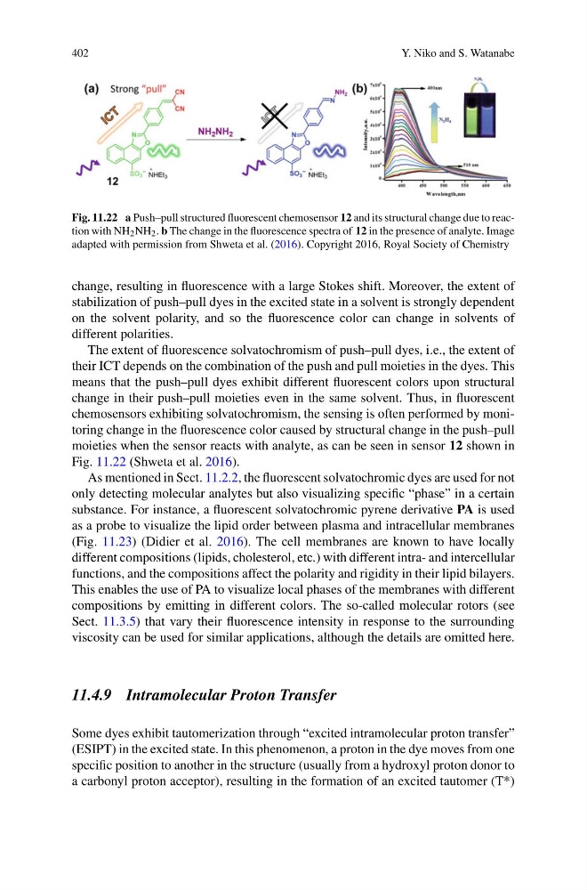
Fig. 11.22 a Push–pull structured fluorescent chemosensor 12 and its structural change due to reaction with NH 2 NH 2 . b The change in the fluorescence spectra of 12 in the presence of analyte. Image
adapted with permission from Shweta et al. (2016). Copyright 2016, Royal Society of Chemistry
change, resulting in fluorescence with a large Stokes shift. Moreover, the extent of
stabilization of push–pull dyes in the excited state in a solvent is strongly dependent
on the solvent polarity, and so the fluorescence color can change in solvents of
different polarities.
The extent of fluorescence solvatochromism of push–pull dyes, i.e., the extent of
their ICT depends on the combination of the push and pull moieties in the dyes. This
means that the push–pull dyes exhibit different fluorescent colors upon structural
change in their push–pull moieties even in the same solvent. Thus, in fluorescent
chemosensors exhibiting solvatochromism, the sensing is often performed by monitoring change in the fluorescence color caused by structural change in the push–pull
moieties when the sensor reacts with analyte, as can be seen in sensor 12 shown in
Fig. 11.22 (Shweta et al. 2016).
As mentioned in Sect. 11.2.2, the fluorescent solvatochromic dyes are used for not
only detecting molecular analytes but also visualizing specific “phase” in a certain
substance. For instance, a fluorescent solvatochromic pyrene derivative PA is used
as a probe to visualize the lipid order between plasma and intracellular membranes
(Fig. 11.23) (Didier et al. 2016). The cell membranes are known to have locally
different compositions (lipids, cholesterol, etc.) with different intra- and intercellular
functions, and the compositions affect the polarity and rigidity in their lipid bilayers.
This enables the use of PA to visualize local phases of the membranes with different
compositions by emitting in different colors. The so-called molecular rotors (see
Sect. 11.3.5) that vary their fluorescence intensity in response to the surrounding
viscosity can be used for similar applications, although the details are omitted here.
11.4.9 Intramolecular Proton Transfer
Some dyes exhibit tautomerization through “excited intramolecular proton transfer”
(ESIPT) in the excited state. In this phenomenon, a proton in the dye moves from one
specific position to another in the structure (usually from a hydroxyl proton donor to
a carbonyl proton acceptor), resulting in the formation of an excited tautomer (T*)
Y. Niko and S. Watanabe
Fig. 11.22 a Push–pull structured fluorescent chemosensor 12 and its structural change due to reaction with NH 2 NH 2 . b The change in the fluorescence spectra of 12 in the presence of analyte. Image
adapted with permission from Shweta et al. (2016). Copyright 2016, Royal Society of Chemistry
change, resulting in fluorescence with a large Stokes shift. Moreover, the extent of
stabilization of push–pull dyes in the excited state in a solvent is strongly dependent
on the solvent polarity, and so the fluorescence color can change in solvents of
different polarities.
The extent of fluorescence solvatochromism of push–pull dyes, i.e., the extent of
their ICT depends on the combination of the push and pull moieties in the dyes. This
means that the push–pull dyes exhibit different fluorescent colors upon structural
change in their push–pull moieties even in the same solvent. Thus, in fluorescent
chemosensors exhibiting solvatochromism, the sensing is often performed by monitoring change in the fluorescence color caused by structural change in the push–pull
moieties when the sensor reacts with analyte, as can be seen in sensor 12 shown in
Fig. 11.22 (Shweta et al. 2016).
As mentioned in Sect. 11.2.2, the fluorescent solvatochromic dyes are used for not
only detecting molecular analytes but also visualizing specific “phase” in a certain
substance. For instance, a fluorescent solvatochromic pyrene derivative PA is used
as a probe to visualize the lipid order between plasma and intracellular membranes
(Fig. 11.23) (Didier et al. 2016). The cell membranes are known to have locally
different compositions (lipids, cholesterol, etc.) with different intra- and intercellular
functions, and the compositions affect the polarity and rigidity in their lipid bilayers.
This enables the use of PA to visualize local phases of the membranes with different
compositions by emitting in different colors. The so-called molecular rotors (see
Sect. 11.3.5) that vary their fluorescence intensity in response to the surrounding
viscosity can be used for similar applications, although the details are omitted here.
11.4.9 Intramolecular Proton Transfer
Some dyes exhibit tautomerization through “excited intramolecular proton transfer”
(ESIPT) in the excited state. In this phenomenon, a proton in the dye moves from one
specific position to another in the structure (usually from a hydroxyl proton donor to
a carbonyl proton acceptor), resulting in the formation of an excited tautomer (T*)