

298
T. Ishi-i
N
S
N
N
Me
39
em 601 nm, F 0.54 (THF/water (1:9, v/v), aggregate)
em 641 nm, F 0.016 (THF/water (4:6, v/v), monomer)
N
S
N
N
N
40
N
S
N
em 617 nm, F 0.34 (THF/water (1:9, v/v), aggregate)
em 671 nm, F 0.02 (THF/water (7:3, v/v), monomer)
N
S
N
N
42
em 621 nm, F 0.20 (water, aggregate),
em 647 nm, F 0.005 (methanol, monomer)
O
O
O
O
Me
Me
6
6
N
Me
Me
N
R
R
43: R = CH 2 CH 2 -O-CO-Ph
F 15.5 GM, 172 GM at 775 nm, em ca. 620 nm, F 0.025 (THF, monomer),
F 101 GM, 217 GM at 775 nm, em ca. 610 nm, F 0.13 (AOT/1-butanol/water, aggregate)
(AOT: anionic surfactant, sodium bis(2-ethylhexyl)sulfosuccinate)
N
N
S
N
N
N
41
em 658 nm, F 0.12 (THF/water (1:9, v/v), aggregate)
em 681 nm, F 0.003 (THF/water (4:6, v/v), monomer)
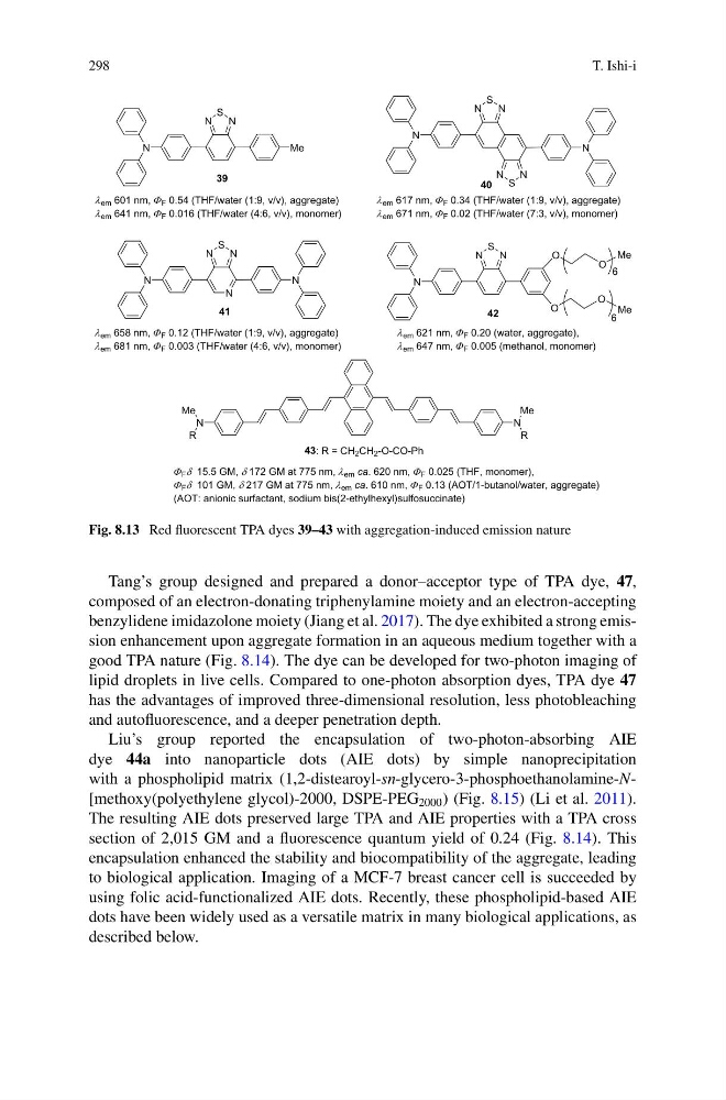
Fig. 8.13 Red fluorescent TPA dyes 39–43 with aggregation-induced emission nature
Tang’s group designed and prepared a donor–acceptor type of TPA dye, 47,
composed of an electron-donating triphenylamine moiety and an electron-accepting
benzylidene imidazolone moiety (Jiang et al. 2017). The dye exhibited a strong emission enhancement upon aggregate formation in an aqueous medium together with a
good TPA nature (Fig. 8.14). The dye can be developed for two-photon imaging of
lipid droplets in live cells. Compared to one-photon absorption dyes, TPA dye 47
has the advantages of improved three-dimensional resolution, less photobleaching
and autofluorescence, and a deeper penetration depth.
Liu’s group reported the encapsulation of two-photon-absorbing AIE
dye 44a into nanoparticle dots (AIE dots) by simple nanoprecipitation
with a phospholipid matrix (1,2-distearoyl-sn-glycero-3-phosphoethanolamine-N[methoxy(polyethylene glycol)-2000, DSPE-PEG 2000 ) (Fig. 8.15) (Li et al. 2011).
The resulting AIE dots preserved large TPA and AIE properties with a TPA cross
section of 2,015 GM and a fluorescence quantum yield of 0.24 (Fig. 8.14). This
encapsulation enhanced the stability and biocompatibility of the aggregate, leading
to biological application. Imaging of a MCF-7 breast cancer cell is succeeded by
using folic acid-functionalized AIE dots. Recently, these phospholipid-based AIE
dots have been widely used as a versatile matrix in many biological applications, as
described below.
T. Ishi-i
N
S
N
N
Me
39
em 601 nm, F 0.54 (THF/water (1:9, v/v), aggregate)
em 641 nm, F 0.016 (THF/water (4:6, v/v), monomer)
N
S
N
N
N
40
N
S
N
em 617 nm, F 0.34 (THF/water (1:9, v/v), aggregate)
em 671 nm, F 0.02 (THF/water (7:3, v/v), monomer)
N
S
N
N
42
em 621 nm, F 0.20 (water, aggregate),
em 647 nm, F 0.005 (methanol, monomer)
O
O
O
O
Me
Me
6
6
N
Me
Me
N
R
R
43: R = CH 2 CH 2 -O-CO-Ph
F 15.5 GM, 172 GM at 775 nm, em ca. 620 nm, F 0.025 (THF, monomer),
F 101 GM, 217 GM at 775 nm, em ca. 610 nm, F 0.13 (AOT/1-butanol/water, aggregate)
(AOT: anionic surfactant, sodium bis(2-ethylhexyl)sulfosuccinate)
N
N
S
N
N
N
41
em 658 nm, F 0.12 (THF/water (1:9, v/v), aggregate)
em 681 nm, F 0.003 (THF/water (4:6, v/v), monomer)
Fig. 8.13 Red fluorescent TPA dyes 39–43 with aggregation-induced emission nature
Tang’s group designed and prepared a donor–acceptor type of TPA dye, 47,
composed of an electron-donating triphenylamine moiety and an electron-accepting
benzylidene imidazolone moiety (Jiang et al. 2017). The dye exhibited a strong emission enhancement upon aggregate formation in an aqueous medium together with a
good TPA nature (Fig. 8.14). The dye can be developed for two-photon imaging of
lipid droplets in live cells. Compared to one-photon absorption dyes, TPA dye 47
has the advantages of improved three-dimensional resolution, less photobleaching
and autofluorescence, and a deeper penetration depth.
Liu’s group reported the encapsulation of two-photon-absorbing AIE
dye 44a into nanoparticle dots (AIE dots) by simple nanoprecipitation
with a phospholipid matrix (1,2-distearoyl-sn-glycero-3-phosphoethanolamine-N[methoxy(polyethylene glycol)-2000, DSPE-PEG 2000 ) (Fig. 8.15) (Li et al. 2011).
The resulting AIE dots preserved large TPA and AIE properties with a TPA cross
section of 2,015 GM and a fluorescence quantum yield of 0.24 (Fig. 8.14). This
encapsulation enhanced the stability and biocompatibility of the aggregate, leading
to biological application. Imaging of a MCF-7 breast cancer cell is succeeded by
using folic acid-functionalized AIE dots. Recently, these phospholipid-based AIE
dots have been widely used as a versatile matrix in many biological applications, as
described below.