

8 Red and Near-IR Fluorescent Two-Photon Absorption Dyes
291
in mitochondria (Sarkar et al. 2016). This dye has a pKa value of 7.95 and exhibits
a yellow to red emission color change in response to pH alterations from 6.0 to
9.0 (Fig. 8.7). Ratiometric two-photon fluorescence microscopy imaging revealed
the heterogeneity of pH values with respect to the specific location of mitochondria
within the cells.
A systematic study of deep-tissue imaging (brain, kidney, liver, lung, and spleen
mouse tissues) by two-photon microscopy using far-red-emitting dyes 16 was
reported by Ahn’s group (Jun et al. 2017). The TPA dye was designed and prepared
based on a π-extended coumarin chromophore that has donor and acceptor moieties
(Fig. 8.7). The intramolecular charge transfer property arising from the donor–
acceptor structure provided the attractive far-red emission and TPA. The two-photon
action cross section was 160 GM, which is comparable to those of the precedent
TPA dyes (e.g., acedan) widely used in two-photon fluorescence microscopy that
emit short-wavelength fluorescence. For deep depth (~150 μm) imaging of kidney
tissue, only the far-red dye 16 can maintain a high signal to background ratio. In
contrast, the ratio decreased in acedan with short-wavelength green emission.
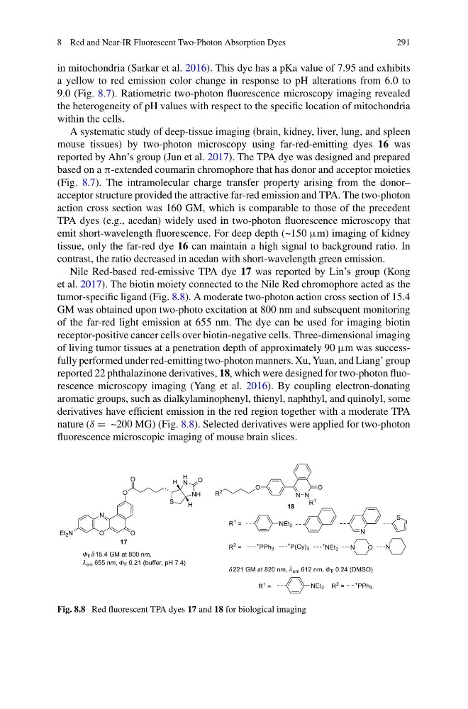
Nile Red-based red-emissive TPA dye 17 was reported by Lin’s group (Kong
et al. 2017). The biotin moiety connected to the Nile Red chromophore acted as the
tumor-specific ligand (Fig. 8.8). A moderate two-photon action cross section of 15.4
GM was obtained upon two-photo excitation at 800 nm and subsequent monitoring
of the far-red light emission at 655 nm. The dye can be used for imaging biotin
receptor-positive cancer cells over biotin-negative cells. Three-dimensional imaging
of living tumor tissues at a penetration depth of approximately 90 μm was successfully performed under red-emitting two-photon manners. Xu, Yuan, and Liang’ group
reported 22 phthalazinone derivatives, 18, which were designed for two-photon fluorescence microscopy imaging (Yang et al. 2016). By coupling electron-donating
aromatic groups, such as dialkylaminophenyl, thienyl, naphthyl, and quinolyl, some
derivatives have efficient emission in the red region together with a moderate TPA
nature (δ = ~200 MG) (Fig. 8.8). Selected derivatives were applied for two-photon
fluorescence microscopic imaging of mouse brain slices.
F 15.4 GM at 800 nm,
em 655 nm, F 0.21 (buffer, pH 7.4)
N
O
Et 2 N
17
O
O
S
O
N
N
O
H
H
H
H
18
221 GM at 820 nm, em 612 nm, F 0.24 (DMSO)
N N
O
O
R
2
R
1
R
1 =
NEt 2
N
S
R
2 =
+ PPh 3
+ P(Cy) 3
+ NEt 3
O
N
N
R
1 =
NEt 2 R
2 =
+ PPh 3
Φ
Φ
Φ
δ
δ
λ
λ
Fig. 8.8 Red fluorescent TPA dyes 17 and 18 for biological imaging
291
in mitochondria (Sarkar et al. 2016). This dye has a pKa value of 7.95 and exhibits
a yellow to red emission color change in response to pH alterations from 6.0 to
9.0 (Fig. 8.7). Ratiometric two-photon fluorescence microscopy imaging revealed
the heterogeneity of pH values with respect to the specific location of mitochondria
within the cells.
A systematic study of deep-tissue imaging (brain, kidney, liver, lung, and spleen
mouse tissues) by two-photon microscopy using far-red-emitting dyes 16 was
reported by Ahn’s group (Jun et al. 2017). The TPA dye was designed and prepared
based on a π-extended coumarin chromophore that has donor and acceptor moieties
(Fig. 8.7). The intramolecular charge transfer property arising from the donor–
acceptor structure provided the attractive far-red emission and TPA. The two-photon
action cross section was 160 GM, which is comparable to those of the precedent
TPA dyes (e.g., acedan) widely used in two-photon fluorescence microscopy that
emit short-wavelength fluorescence. For deep depth (~150 μm) imaging of kidney
tissue, only the far-red dye 16 can maintain a high signal to background ratio. In
contrast, the ratio decreased in acedan with short-wavelength green emission.
Nile Red-based red-emissive TPA dye 17 was reported by Lin’s group (Kong
et al. 2017). The biotin moiety connected to the Nile Red chromophore acted as the
tumor-specific ligand (Fig. 8.8). A moderate two-photon action cross section of 15.4
GM was obtained upon two-photo excitation at 800 nm and subsequent monitoring
of the far-red light emission at 655 nm. The dye can be used for imaging biotin
receptor-positive cancer cells over biotin-negative cells. Three-dimensional imaging
of living tumor tissues at a penetration depth of approximately 90 μm was successfully performed under red-emitting two-photon manners. Xu, Yuan, and Liang’ group
reported 22 phthalazinone derivatives, 18, which were designed for two-photon fluorescence microscopy imaging (Yang et al. 2016). By coupling electron-donating
aromatic groups, such as dialkylaminophenyl, thienyl, naphthyl, and quinolyl, some
derivatives have efficient emission in the red region together with a moderate TPA
nature (δ = ~200 MG) (Fig. 8.8). Selected derivatives were applied for two-photon
fluorescence microscopic imaging of mouse brain slices.
F 15.4 GM at 800 nm,
em 655 nm, F 0.21 (buffer, pH 7.4)
N
O
Et 2 N
17
O
O
S
O
N
N
O
H
H
H
H
18
221 GM at 820 nm, em 612 nm, F 0.24 (DMSO)
N N
O
O
R
2
R
1
R
1 =
NEt 2
N
S
R
2 =
+ PPh 3
+ P(Cy) 3
+ NEt 3
O
N
N
R
1 =
NEt 2 R
2 =
+ PPh 3
Φ
Φ
Φ
δ
δ
λ
λ
Fig. 8.8 Red fluorescent TPA dyes 17 and 18 for biological imaging