

6 Molecular and Crystal Structures of Polymorphic …
251
(a) 1
(b) 2
(c) 3
(d) 4
(e) 5
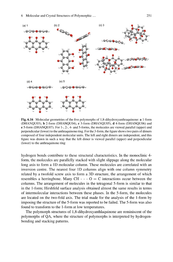
Fig. 6.16 Molecular geometries of the five polymorphs of 1,8-dihydroxyanthraquinone: a 1-form
(DHANQU03), b 2-form (DHANQU04), c 3-form (DHANQU05), d 4-form (DHANQU06) and
e 5-form (DHANQU07). For 1-, 2-, 4- and 5-forms, the molecules are viewed parallel (upper) and
perpendicular (lower) to the anthraquinone ring. For the 3-form, the figure shows two pairs of dimers
composed of four independent molecular units. The left and right dimers are independent, and this
figure was drawn in such a way that the left dimer is viewed parallel (upper) and perpendicular
(lower) to the anthraquinone ring
hydrogen bonds contribute to these structural characteristics. In the monoclinic 4form, the molecules are parallelly stacked with slight slippage along the molecular
long axis to form a 1D molecular column. These molecules are correlated with an
inversion centre. The nearest four 1D columns align with one column symmetry
related by a twofold screw axis to form a 3D structure, the arrangement of which
resembles a herringbone. Many CH - - - O = C interactions occur between the
columns. The arrangement of molecules in the tetragonal 5-form is similar to that
in the 1-form; Hirshfeld surface analysis obtained almost the same results in terms
of intermolecular interactions between these phases. In the 5-form, the molecules
are located on the two-fold axis. The trial made for the analysis of the 1-form by
imposing the structure of the 5-form was reported to be failed. The 5-form was also
found to transform to the 1-form at low temperatures.
The polymorph structures of 1,8-dihydroxyanthlaquinone are reminiscent of the
polymorphs of QA, where the structure of polymorphs is interpreted by hydrogenbonding and stacking patterns.
251
(a) 1
(b) 2
(c) 3
(d) 4
(e) 5
Fig. 6.16 Molecular geometries of the five polymorphs of 1,8-dihydroxyanthraquinone: a 1-form
(DHANQU03), b 2-form (DHANQU04), c 3-form (DHANQU05), d 4-form (DHANQU06) and
e 5-form (DHANQU07). For 1-, 2-, 4- and 5-forms, the molecules are viewed parallel (upper) and
perpendicular (lower) to the anthraquinone ring. For the 3-form, the figure shows two pairs of dimers
composed of four independent molecular units. The left and right dimers are independent, and this
figure was drawn in such a way that the left dimer is viewed parallel (upper) and perpendicular
(lower) to the anthraquinone ring
hydrogen bonds contribute to these structural characteristics. In the monoclinic 4form, the molecules are parallelly stacked with slight slippage along the molecular
long axis to form a 1D molecular column. These molecules are correlated with an
inversion centre. The nearest four 1D columns align with one column symmetry
related by a twofold screw axis to form a 3D structure, the arrangement of which
resembles a herringbone. Many CH - - - O = C interactions occur between the
columns. The arrangement of molecules in the tetragonal 5-form is similar to that
in the 1-form; Hirshfeld surface analysis obtained almost the same results in terms
of intermolecular interactions between these phases. In the 5-form, the molecules
are located on the two-fold axis. The trial made for the analysis of the 1-form by
imposing the structure of the 5-form was reported to be failed. The 5-form was also
found to transform to the 1-form at low temperatures.
The polymorph structures of 1,8-dihydroxyanthlaquinone are reminiscent of the
polymorphs of QA, where the structure of polymorphs is interpreted by hydrogenbonding and stacking patterns.