

250
S. Matsumoto and J. Hwang
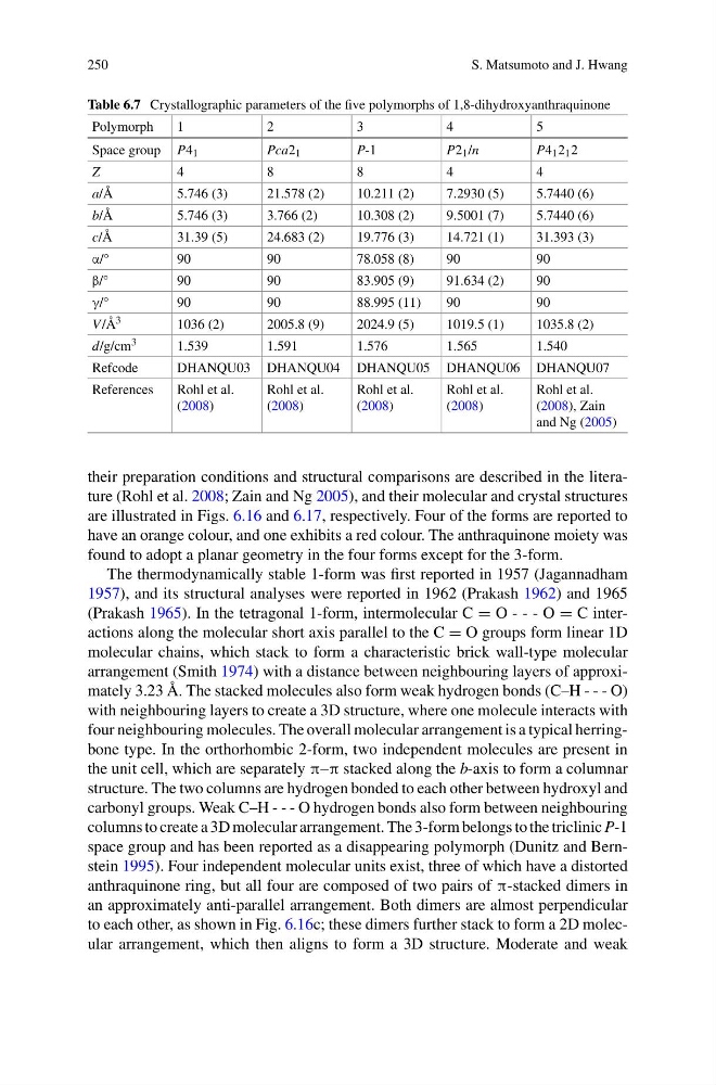
Table 6.7 Crystallographic parameters of the five polymorphs of 1,8-dihydroxyanthraquinone
Polymorph
1
2
3
4
5
Space group P4 1
Pca2 1
P-1
P2 1 /n
P4 1 2 1 2
Z
4
8
8
4
4
a/Å
5.746 (3)
21.578 (2)
10.211 (2)
7.2930 (5)
5.7440 (6)
b/Å
5.746 (3)
3.766 (2)
10.308 (2)
9.5001 (7)
5.7440 (6)
c/Å
31.39 (5)
24.683 (2)
19.776 (3)
14.721 (1)
31.393 (3)
α/°
90
90
78.058 (8)
90
90
β/°
90
90
83.905 (9)
91.634 (2)
90
γ/°
90
90
88.995 (11)
90
90
V /Å 3
1036 (2)
2005.8 (9)
2024.9 (5)
1019.5 (1)
1035.8 (2)
d/g/cm 3
1.539
1.591
1.576
1.565
1.540
Refcode
DHANQU03 DHANQU04 DHANQU05 DHANQU06 DHANQU07
References
Rohl et al.
(2008)
Rohl et al.
(2008)
Rohl et al.
(2008)
Rohl et al.
(2008)
Rohl et al.
(2008), Zain
and Ng (2005)
their preparation conditions and structural comparisons are described in the literature (Rohl et al. 2008; Zain and Ng 2005), and their molecular and crystal structures
are illustrated in Figs. 6.16 and 6.17, respectively. Four of the forms are reported to
have an orange colour, and one exhibits a red colour. The anthraquinone moiety was
found to adopt a planar geometry in the four forms except for the 3-form.
The thermodynamically stable 1-form was first reported in 1957 (Jagannadham
1957), and its structural analyses were reported in 1962 (Prakash 1962) and 1965
(Prakash 1965). In the tetragonal 1-form, intermolecular C = O - - - O = C interactions along the molecular short axis parallel to the C = O groups form linear 1D
molecular chains, which stack to form a characteristic brick wall-type molecular
arrangement (Smith 1974) with a distance between neighbouring layers of approximately 3.23 Å. The stacked molecules also form weak hydrogen bonds (C–H - - - O)
with neighbouring layers to create a 3D structure, where one molecule interacts with
four neighbouring molecules. The overall molecular arrangement is a typical herringbone type. In the orthorhombic 2-form, two independent molecules are present in
the unit cell, which are separately π–π stacked along the b-axis to form a columnar
structure. The two columns are hydrogen bonded to each other between hydroxyl and
carbonyl groups. Weak C–H - - - O hydrogen bonds also form between neighbouring
columns to create a 3D molecular arrangement. The 3-form belongs to the triclinic P-1
space group and has been reported as a disappearing polymorph (Dunitz and Bernstein 1995). Four independent molecular units exist, three of which have a distorted
anthraquinone ring, but all four are composed of two pairs of π-stacked dimers in
an approximately anti-parallel arrangement. Both dimers are almost perpendicular
to each other, as shown in Fig. 6.16c; these dimers further stack to form a 2D molecular arrangement, which then aligns to form a 3D structure. Moderate and weak
S. Matsumoto and J. Hwang
Table 6.7 Crystallographic parameters of the five polymorphs of 1,8-dihydroxyanthraquinone
Polymorph
1
2
3
4
5
Space group P4 1
Pca2 1
P-1
P2 1 /n
P4 1 2 1 2
Z
4
8
8
4
4
a/Å
5.746 (3)
21.578 (2)
10.211 (2)
7.2930 (5)
5.7440 (6)
b/Å
5.746 (3)
3.766 (2)
10.308 (2)
9.5001 (7)
5.7440 (6)
c/Å
31.39 (5)
24.683 (2)
19.776 (3)
14.721 (1)
31.393 (3)
α/°
90
90
78.058 (8)
90
90
β/°
90
90
83.905 (9)
91.634 (2)
90
γ/°
90
90
88.995 (11)
90
90
V /Å 3
1036 (2)
2005.8 (9)
2024.9 (5)
1019.5 (1)
1035.8 (2)
d/g/cm 3
1.539
1.591
1.576
1.565
1.540
Refcode
DHANQU03 DHANQU04 DHANQU05 DHANQU06 DHANQU07
References
Rohl et al.
(2008)
Rohl et al.
(2008)
Rohl et al.
(2008)
Rohl et al.
(2008)
Rohl et al.
(2008), Zain
and Ng (2005)
their preparation conditions and structural comparisons are described in the literature (Rohl et al. 2008; Zain and Ng 2005), and their molecular and crystal structures
are illustrated in Figs. 6.16 and 6.17, respectively. Four of the forms are reported to
have an orange colour, and one exhibits a red colour. The anthraquinone moiety was
found to adopt a planar geometry in the four forms except for the 3-form.
The thermodynamically stable 1-form was first reported in 1957 (Jagannadham
1957), and its structural analyses were reported in 1962 (Prakash 1962) and 1965
(Prakash 1965). In the tetragonal 1-form, intermolecular C = O - - - O = C interactions along the molecular short axis parallel to the C = O groups form linear 1D
molecular chains, which stack to form a characteristic brick wall-type molecular
arrangement (Smith 1974) with a distance between neighbouring layers of approximately 3.23 Å. The stacked molecules also form weak hydrogen bonds (C–H - - - O)
with neighbouring layers to create a 3D structure, where one molecule interacts with
four neighbouring molecules. The overall molecular arrangement is a typical herringbone type. In the orthorhombic 2-form, two independent molecules are present in
the unit cell, which are separately π–π stacked along the b-axis to form a columnar
structure. The two columns are hydrogen bonded to each other between hydroxyl and
carbonyl groups. Weak C–H - - - O hydrogen bonds also form between neighbouring
columns to create a 3D molecular arrangement. The 3-form belongs to the triclinic P-1
space group and has been reported as a disappearing polymorph (Dunitz and Bernstein 1995). Four independent molecular units exist, three of which have a distorted
anthraquinone ring, but all four are composed of two pairs of π-stacked dimers in
an approximately anti-parallel arrangement. Both dimers are almost perpendicular
to each other, as shown in Fig. 6.16c; these dimers further stack to form a 2D molecular arrangement, which then aligns to form a 3D structure. Moderate and weak