

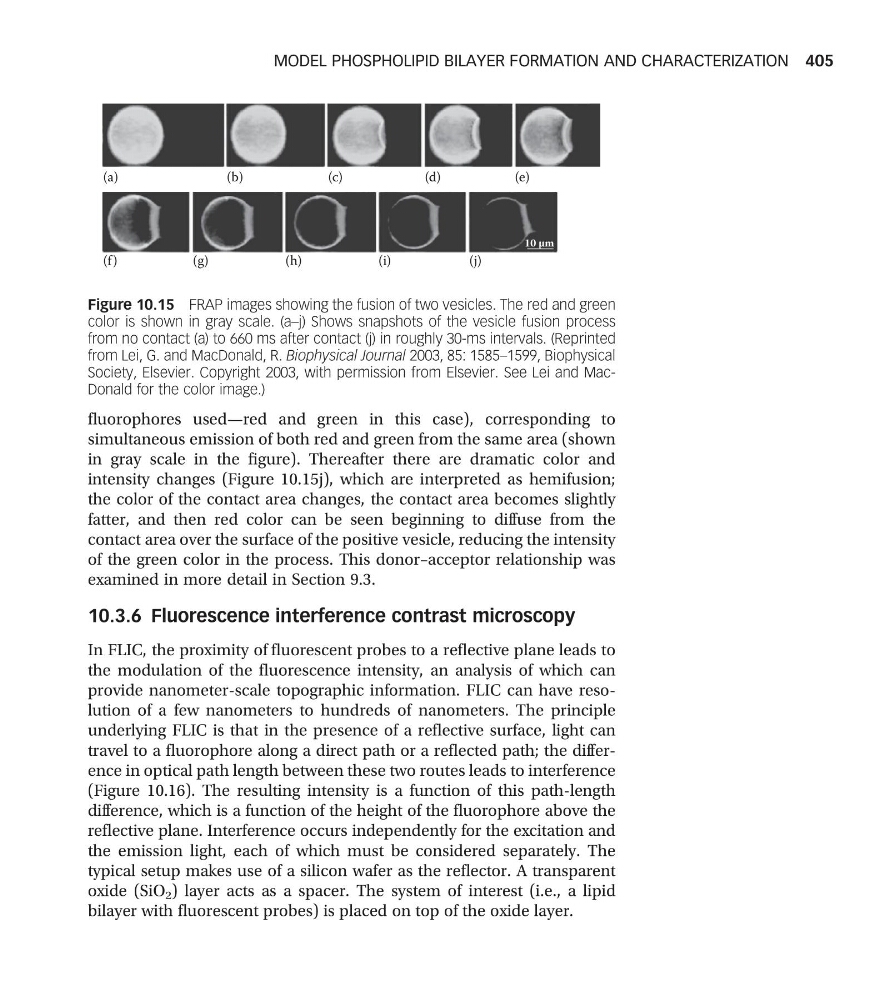
fluorophores used—red and green in this case), corresponding to
simultaneous emission of both red and green from the same area (shown
in gray scale in the figure). Thereafter there are dramatic color and
intensity changes (Figure 10.15j), which are interpreted as hemifusion;
the color of the contact area changes, the contact area becomes slightly
fatter, and then red color can be seen beginning to diffuse from the
contact area over the surface of the positive vesicle, reducing the intensity
of the green color in the process. This donor–acceptor relationship was
examined in more detail in Section 9.3.
10.3.6 Fluorescence interference contrast microscopy
In FLIC, the proximity of fluorescent probes to a reflective plane leads to
the modulation of the fluorescence intensity, an analysis of which can
provide nanometer-scale topographic information. FLIC can have resolution of a few nanometers to hundreds of nanometers. The principle
underlying FLIC is that in the presence of a reflective surface, light can
travel to a fluorophore along a direct path or a reflected path; the difference in optical path length between these two routes leads to interference
(Figure 10.16). The resulting intensity is a function of this path-length
difference, which is a function of the height of the fluorophore above the
reflective plane. Interference occurs independently for the excitation and
the emission light, each of which must be considered separately. The
typical setup makes use of a silicon wafer as the reflector. A transparent
oxide (SiO 2 ) layer acts as a spacer. The system of interest (i.e., a lipid
bilayer with fluorescent probes) is placed on top of the oxide layer.
10 µm
(a)
(f )
(g)
(h)
(i)
(j)
(b)
(c)
(d)
(e)
Figure 10.15 FRAP images showing the fusion of two vesicles. The red and green
color is shown in gray scale. (a–j) Shows snapshots of the vesicle fusion process
from no contact (a) to 660 ms after contact (j) in roughly 30-ms intervals. (Reprinted
from Lei, G. and MacDonald, R. Biophysical Journal 2003, 85: 1585–1599, Biophysical
Society, Elsevier. Copyright 2003, with permission from Elsevier. See Lei and MacDonald for the color image.)
MODEL PHOSPHOLIPID BILAYER FORMATION AND CHARACTERIZATION 405
simultaneous emission of both red and green from the same area (shown
in gray scale in the figure). Thereafter there are dramatic color and
intensity changes (Figure 10.15j), which are interpreted as hemifusion;
the color of the contact area changes, the contact area becomes slightly
fatter, and then red color can be seen beginning to diffuse from the
contact area over the surface of the positive vesicle, reducing the intensity
of the green color in the process. This donor–acceptor relationship was
examined in more detail in Section 9.3.
10.3.6 Fluorescence interference contrast microscopy
In FLIC, the proximity of fluorescent probes to a reflective plane leads to
the modulation of the fluorescence intensity, an analysis of which can
provide nanometer-scale topographic information. FLIC can have resolution of a few nanometers to hundreds of nanometers. The principle
underlying FLIC is that in the presence of a reflective surface, light can
travel to a fluorophore along a direct path or a reflected path; the difference in optical path length between these two routes leads to interference
(Figure 10.16). The resulting intensity is a function of this path-length
difference, which is a function of the height of the fluorophore above the
reflective plane. Interference occurs independently for the excitation and
the emission light, each of which must be considered separately. The
typical setup makes use of a silicon wafer as the reflector. A transparent
oxide (SiO 2 ) layer acts as a spacer. The system of interest (i.e., a lipid
bilayer with fluorescent probes) is placed on top of the oxide layer.
10 µm
(a)
(f )
(g)
(h)
(i)
(j)
(b)
(c)
(d)
(e)
Figure 10.15 FRAP images showing the fusion of two vesicles. The red and green
color is shown in gray scale. (a–j) Shows snapshots of the vesicle fusion process
from no contact (a) to 660 ms after contact (j) in roughly 30-ms intervals. (Reprinted
from Lei, G. and MacDonald, R. Biophysical Journal 2003, 85: 1585–1599, Biophysical
Society, Elsevier. Copyright 2003, with permission from Elsevier. See Lei and MacDonald for the color image.)
MODEL PHOSPHOLIPID BILAYER FORMATION AND CHARACTERIZATION 405